
The Common Vein Copyright 2010
Introduction
Normal
Nodule Vascularity
There are 3 types of vascularity in thyroid nodules;
Type 1 – peripheral vascularity or no vascularity and it reflects benign disease
Type 2 – small amount of internal vascularity in the lesion and a little more concerning
Type 3 – very vascular throughout lesion is the most worrisome and suspicious for malignancy
However many benign lesions have type 3 vascularity and most papillary carcinomas are avascular.
Therefore vascularity should only be used as an adjunctive sign – ie concern should be raised if there is type 3 vascularity and the lesion is otherwise indeterminate.
Type 1
Multiplae Benign Appearing Nodules – Peripheral Doppler Flow |
|
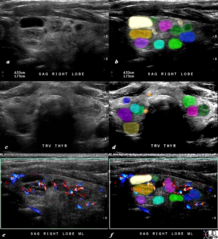
The 80 year old male presents with an asymptomatic multinodular goiter, consisting many nodules of varying size and echogenocity, as seen by sagittal ultrasound (a,b) and transverse imaging images (c,d), and Doppler imaging (e,f). The multiple nodules that are not border forming are within the confines of the parenchyma and do not alter the shape of the gland. The right lobe of the thyroid measures about 4.5 cms in sagittal 1.5cms.in A-P dimension, and 1.5cms in the transverse plane. The gland is therefore not enlarged. In addition the gland has a normal appearance in the transverse projection and the borders are not rounded to suggest enlargement. The Doppler study shows no internal vascularity in any of the nodules visualized. These findings are consistent with a non toxic multinodular thyroid gland, and not truly a goiter since the gland is not enlarged. Courtesy Ashley Davidoff MD Copyright 2010 94780c04b02.8s |
Type 2
Type 2 – Small Amount of Internal Vascularity of PApillary Carcinoma |
|
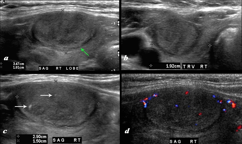
A large nodule in the thyroid occupies almost the entire right lobe (a). The nodule measures 2.9cms by 1.5cms. The gland is not enlarged and measures 3.5cms (craniocaudad), by 1.8cms (A-P) by 2.4cms (transverse). The nodule is almost isoechoic with normal thyroid but shows internal irregular areas of hypoechogenicity, regions of isoechogenicity, as well as microcalcifications (white arrows (c). There is irregularity of the border at the posterior aspect of the nodule green arrow a). The halo shows irregular borders in this region as well. Internal vascularity is minimal (d). The irregular surface is concerning for a malignant processes. The diagnosis in this patient was papillary carcinoma Courtesy Ashley Davidoff MD Copyright 2010 74909c02L.8 |
Type 3
Type 3 – Follicular Adenoma |
|
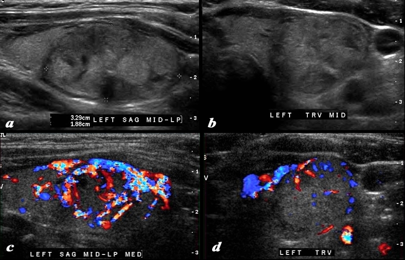
This 37 year old female presents with single nodule in the left lobe of the thyroid. Ultrasound of the mass in sagittal (a) and transverse view (b) reveals a complex mass with isoechoic and hypoechoic nodular components. The normal thyroid tissue is seen superiorly in image a and medically in image b. In the sagittal view there also appears to be a small shadowing macrocalcification (arrow). The halo is seen partly around the superior aspect on the sagittal view and shows no definite irregularity. The Doppler study shows mostly a peripheral pattern but some prominent central vessels are shown. There are no definite criteria to suggest malignant disease but in view of the size, age and vascular pattern biopsy was performed and showed a follicular adenoma. c Courtesy Ashley Davidoff MD Copyright 2010 95654c02.8 |
Type 3 Vascularity – Medullary Carcinoma |
|
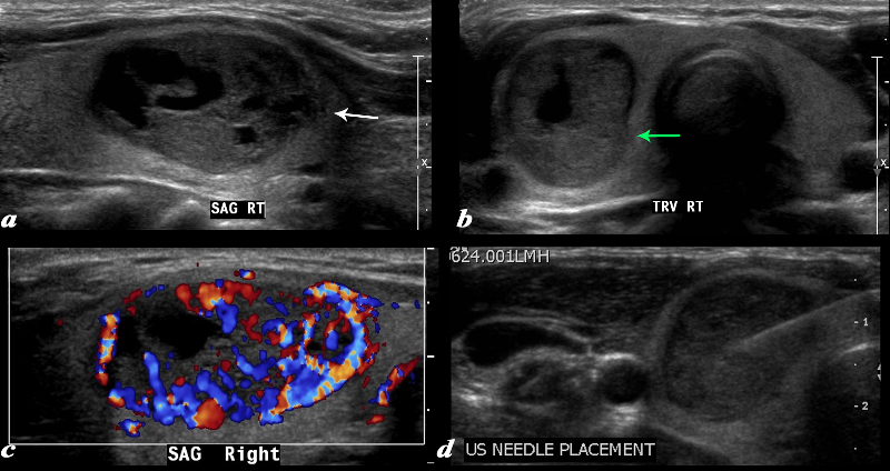
A large complex mass occupies more than half the right lobe of the thyroid gland is seen by ultrasound. The mass measures 2.6cms in sagittal and in A-P dimension measures 1.7cms(a). In transverse it measure 1.7cms. It is characterized by a mildly hypoechoic matrix with cystic type serpiginous components. (a) The margins are irregular (white arrow, a) There is an incomplete halo which shows regions of irregularity (green arrow) The lesion is extremely vascular both peripherally and centrally (c) A biopsy was performed and showed medullary carcinoma. Barry Sacks MD Copyright 2010 97339cL01.8 |
Thyroiditis
Thyroiditis Diffuse Increase in Vacscularity |
|
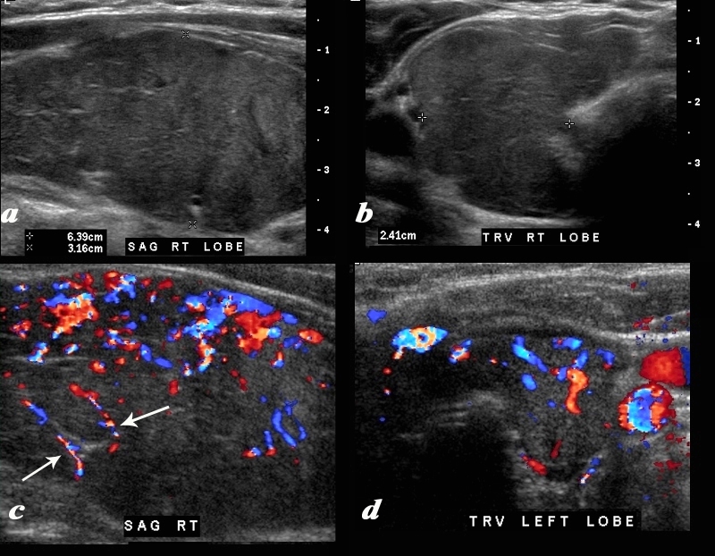
A diffusely enlarged, heterogeneous thyroid gland is seen in this 30 year old hypothyroid female patient. The thyroid measures 6.4cms (craniocaudad), by 3.2cms (A-P) by 2.4cms (transverse). Clinical findings were consistent with thyroiditis, with biochemical findings suggesting hypothyroidism. The sagittal view shows coarse heterogeneous echo texture with fine white bands consistent with fibrosis. The increased vascularity is seen throughout the gland (c,d), but is also seen particularly along some of the bands in the posterior aspect of the gland (c arrows). The enlarged gland in the transverse dimension is almost round. (b). This rounded shape in the transverse dimension is a clue to the presence of the enlarged gland, even before the measurements are taken and evaluated. The findings are consistent with a thyroiditis. Courtesy Ashley Davidoff MD Copyright 2010 94583c01.8 |