
The Common Vein Copyright 2010
Definition
Medullary Carcinoma is a primary malignant neoplastic tumor of the parafollicular C cells in the thyroid gland. Medullary carcinoma is the 3rd most common primary cancer of the thyroid.
Medullary Carcinoma is caused by sporadic malignant transformation in 75 % of cases, and by inherited genetic mutations in the remaining 25% of cases. Familial (inherited) types include Multiple Endocrine Neoplasias (MEN 2A and 2B), as well as non-MEN medullary carcinoma.
Genetic mutations in parafollicular C cells results in unchecked proliferation and mass production of calcitonin. If untreated, medullary carcinoma can cause local mass effect and metastasize to distant organs.
Structural changes of medullary carcinoma involve formation of a firm mass within the thyroid gland. Histologically, amyloid material can be seen from overproduction of the pro-hormone of calcitonin.=
Functional changes of medullary carcinoma include increased production and release of calcitonin by the parafollicular C cells. Lab tests will show elevated levels of calcitonin, with levels greater than 100 pg/ml having a 100% positive predictive value for medullary thyroid carcinoma. There is typically no change in thyroid hormone production or release.
Clinically, medullary carcinoma often presents as a firm thyroid nodule. More extensive disease can present with distant metastases, dysphagia, hoarseness, cervical lymphadenopathy, and/or diarrhea from the increased secretory properties of high levels of calcitonin. In patients with MEN syndromes, the patients can present with other endocrine tumors simultaneously.
Imaging can be performed with ultrasound and/or CT scan to assess for metastatic disease. Ultrasound can guide fine needle biopsy as well as identify lymph node involvement. Diagnosis is best made with biopsy of the nodule.
Treatment of medullary carcinoma is mainly surgical. Total thyroidectomy and neck dissection, if indicated, is the first line of treatment. Clinicians should follow up with the patients and rule out a familial cause of the cancer, such as MEN syndrome.
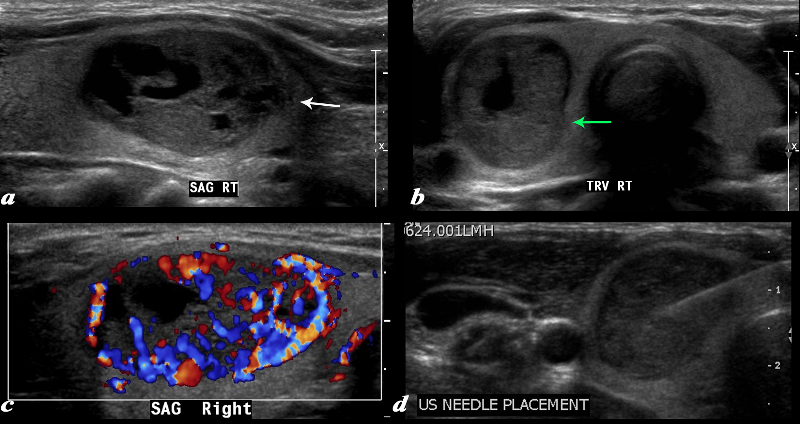
Ultrasound – Medullary Carcinoma |
|
A large complex mass occupies more than half the right lobe of the thyroid gland is seen by ultrasound. The mass measures 2.6cms in sagittal and in A-P dimension measures 1.7cms(a). In transverse it measure 1.7cms. It is characterized by a mildly hypoechoic matrix with cystic type serpiginous components. (a) The margins are irregular (white arrow, a) There is an incomplete halo which shows regions of irregularity (green arrow) The lesion is extremely vascular both peripherally and centrally (c) A biopsy was performed and showed medullary carcinoma. Courtesy Barry Sacks MD Copyright 2010 97339cL01.8 |
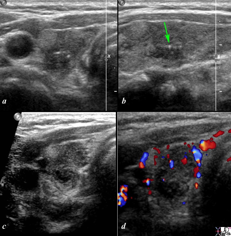
Medullary Carcinoma |
|
This is a thyroid scan from a 53 year old female with a clinical history of bilateral thyroid nodules. The scan shows a heterogeneous nodule in the mid to upper pole of the right lobe that is ill defined both on the transverse view (a) as well as the sagittal view (b). An echogenic nodule is part of the complex lesion. In addition non-shadowing microcalcifications with ring down (green arrow) are seen in both the transverse view (a) and sagittal view (b). On magnification and more contrasted settings the heterogeneity of the lesion is further demonstrated and low vascularity is shown on the Doppler interrogation (d). The pathological diagnosis was medullary cell carcinoma. Courtesy Barry Sacks MD Copyright 2010 99389cL.8s |
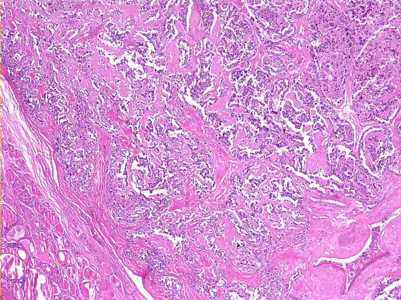
Nests of Hyperchromatic cells Amyloid in Stroma 4X H&E |
|
The histological section at 4X magnification using H&E stain shows findings consistent with medullary thyroid carcinoma characterized by solid nest of hyperchromatic cells with eosinophilic material which represents amyloid in the stroma Image Courtesy Ashraf Khan MD. Department of Pathology, University of Massachusetts Medical School. 99406.8 |
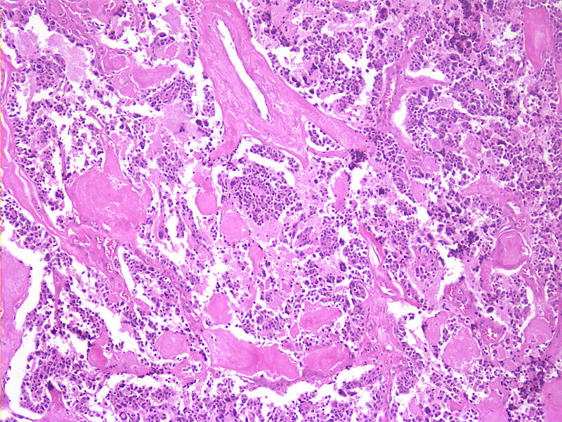
Nests of Hyperchromatic cells Amyloid in Stroma 4X H&E |
|
The histological section at 20X magnification using H&E stain shows findings consistent with medullary thyroid carcinoma characterized by solid nest of hyperchromatic cells with eosinophilic material which represents amyloid in the stroma Image Courtesy Ashraf Khan MD. Department of Pathology, University of Massachusetts Medical School. 99405.8 |