
The Common Vein Copyright 2010
Introduction
Ultrasound Guided Procedures
Therapies
Large cystic lesions
Aspiration
Sclerosis (Doxycycline vs Alcohol)
Ablation
Hyperfunctioning nodules
Large benign nodules to decrease bulk
Metastatic papillary carcinoma nodes
? for Primary neoplastic lesions? Anaplastic Lesions
Techniques
Percutaneous alcohol injection
Laser photocoagulation
?RF and cryotherapy
?Therapeutic Ultrasound
Lymphoma Chemotherapy and XRT
|
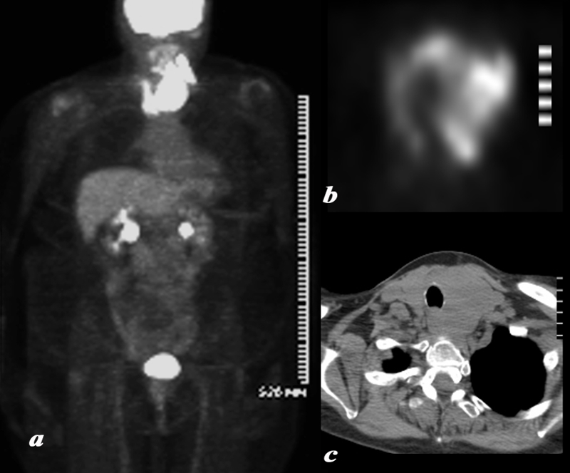
Lymphoma of the Thyroid |
|
A large and very PET avid left lobe of the thyroid is demonstrated on the body PET scan shown in full body view in (a) and in transverse view of the thyroid in (b). The patient is a 62 year female who had a biopsy proven lymphoma of the thyroid.. The CT shows asymmetric and smooth and homogeneous enlargement of the thyroid. The findings are consistent with the final biopsy proven diagnosis of lymphoma. Courtesy Ashley Davidoff MD Copyright 2010 95855c01.8 |
|
PET following Chemotherapy and Radiation Therapy |
|
The patient is a 62 year female who had a biopsy proven lymphoma of the thyroid . A large and very PET avid left lobe of the thyroid is demonstrated on the body PET scan shown in full body view in (a) with resolution of the mass and significant and likely remission changes (b) following chemotherapy and radiotherapy. At the base of the neck there is a U shaped pattern corresponding to residual thyroid tissue. The SUV was 5, higher than typically expected which usually ranges between 1and 2 suggesting persistent moderate uptake. This is likely due to radiation thyroiditis. The findings are most consistent with lymphoma in remission following therapy and likely associated XRt thyroiditis. Courtesy Ashley Davidoff MD Copyright 2010 95855c02.8 |
