The Common Vein Copyright 2010
Definition
Grave’s disease is an autoimmune disease of the thyroid gland usually resulting in goiter and thyrotoxicosis.
It is caused by antibodies against the TSH receptor in the thyroid cell, which cause the thyroid to increase in size.
Graves’ disease results in symptoms of hyperthyroidism, together with an enlarged thyroid glad, as well as the possible accompanying effects of exophthalmos and dermopathy.
Structurally Graves’ disease is characterized by a diffusely enlarged thyroid gland.
Functional changes include a hypermetabolic state induced by circulating thyroid hormone excess, as well as eye findings and skin findings in affected people.
Clinically, most common symptoms include the general findings of an overactive thyroid state: palpitations, tremor, nervousness, fatigue and weakness, weight loss, diarrhea, excess sweating, and heat intolerance. The eye findings of Graves’ disease include upper lid retraction and stare and lid lag in milder forms; proptosis and extroocular muscle involvement in more advanced stages. The characteristic skin finding, pretibial myxedema, is rare. It consists of thickening of the skin, particularly over the lower tibia.
Labs of Graves’ disease are a suppressed TSH together with an elevation in free T4 and/or T3. Thyroid autoantibodies are often present.
The most helpful imaging procedure for suspected Graves’ disease is a radioactive scan and uptake. The scan may help clarify the size of the gland and the iodine uptake confirms the diagnosis when it is increased and diffuse.
Treatment of Graves diase is primarily directed at managing the hyperthryoidism. Antithyroid drugs are helpful, especially in young people with mild disease and smaller glands. Spontaneous remission occurs in a significant percentage within several years, but relapse is high. Radioactive iodine therapy with 131I is the preferred treatment for most adults with Graves’ disease. sometimes patients are treated for a period of time with medicine to achieve control of hyperthyroidism before the RAI is given. This treatment results in permanent hypothyroidism in the majority of patients, who are then treated with thyroxine replacement. Finally, surgery as subtotal thyroidectomy is indicated in some patients with either very large glands or resistant disease.
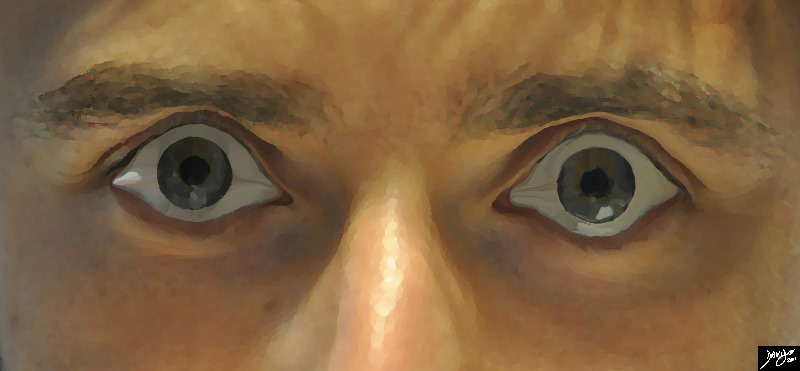
Exopthalmos Note the Whites of the Eyes |
|
Bilateral exopthalmos or bulging of the eyes is most commonly associated with Grave’s disease which is an autoimmune disease associated with thyrotoxicosis. Upperlid retraction results in the ability to see the whites of the eyes on both sides of the iris. Courtesy Ashley Davidoff MD Copyright 2011 100169pb03b.8s |
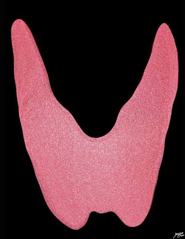
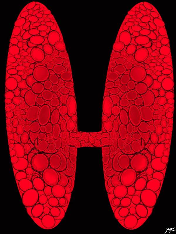
Diffuse Enlargement in Graves Disease |
|
Patients with Graves disease have diffuse enlargement of a hyperfunctioning thyroid gland. Courtesy Ashley DAvidoff MD copyright 2010 all rights reserved 93852.d03.8s |
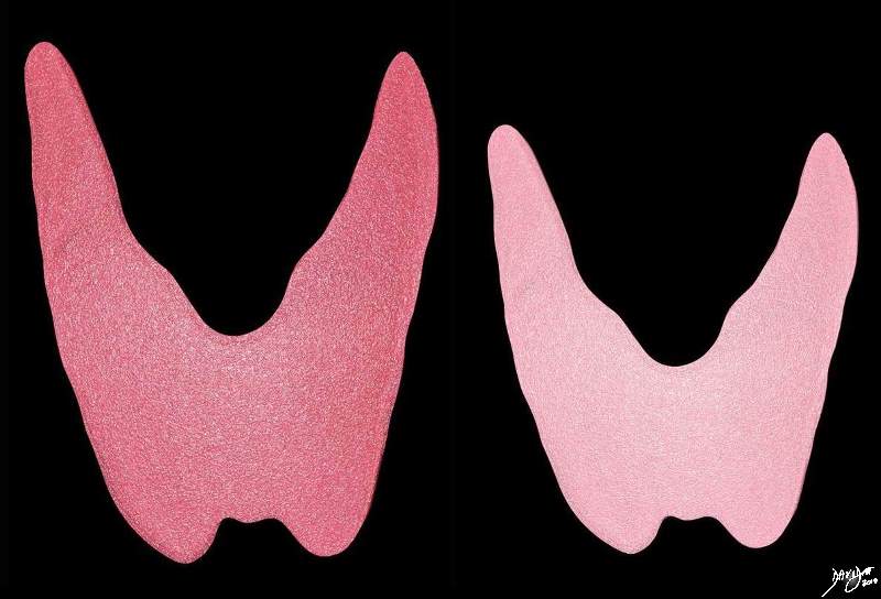
Graves Disease vs Normal Thyroid |
|
Patients with Graves’ disease have diffuse enlargement of a hyperfunctioning gland as seen in the right image (red). The left image (pink) is a normal gland. Ashley DAvidoff MD copyright 2010 all rights reserved 93852.d03c.8s |
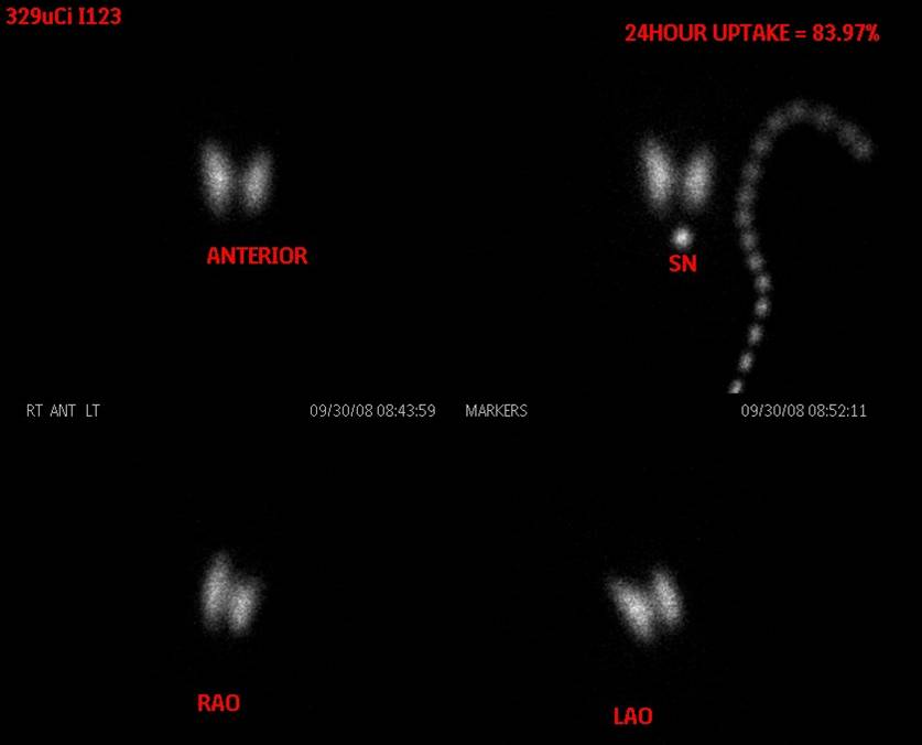
Graves Disease 24hour Uptake Excessively High |
|
This thyroid scan shows iodine uptake of 84% at 24 hours, which is diagnostic of Graves’ disease Image Courtesy Barry Sacks MD 97220 |
|
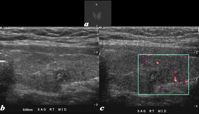
Grave’s Disease and Thyrotoxicosis |
|
This series includes a nuclear medicine thyroid scan, and an ultrasound of a patient with Grave’s disease and a small papillary carcinoma. Image(a) is a rather small nuclear medicine thyroid scan and shows diffuse increase in uptake of the radioisotope in a enlarged gland, confirming the underlying clinically apparent Graves disease. Image b is a sagittal view of the right lobe of the thyroid and shows a 6mm hypoechoic solid lesion with microcalcifications. Image c shows hypovascularity of the lesion. A biopsy was performed and papillary thyroid carcinoma in a patient with Graves’ disease was diagnosed. Courtesy Barry Sacks MD Copyright 2010 97025cL.8 |
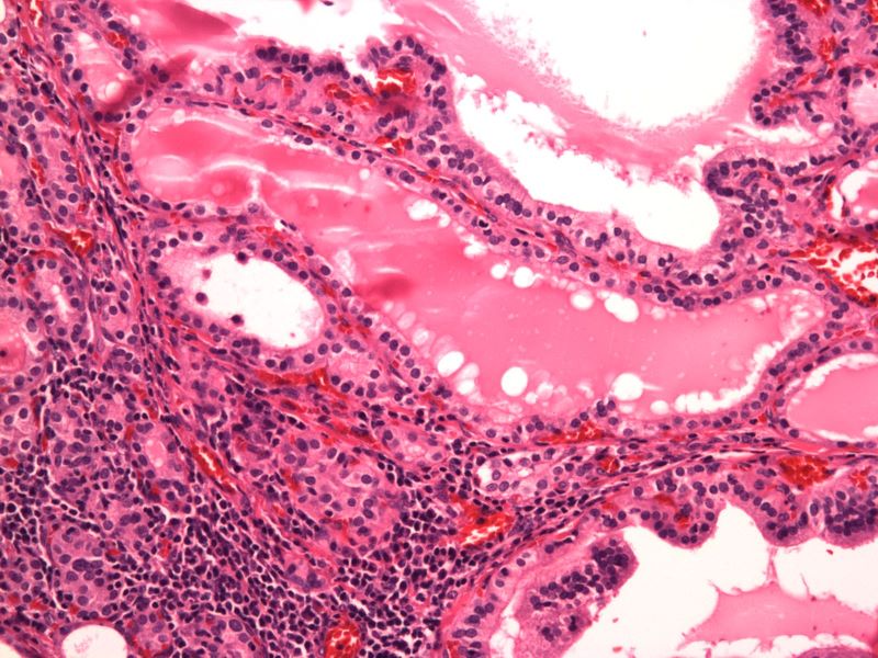
Tall Columnar Cells, Scalloping of Colloid, Lymphoid Infiltrate 20X H&E |
|
The histological section at 20X magnification using H&E stain shows follicles lined by tall columnar cells with scalloping of the colloid at the edges of the follicle and lymphoid infiltrate in the stroma Image Courtesy Ashraf Khan MD. Department of Pathology, University of Massachusetts Medical School 99399.8 |
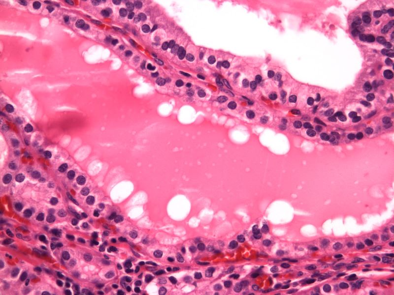
Tall Columnar Cells, Scalloping of Colloid, Lymphoid Infiltrate 20X H&E |
|
The histological section at 40X magnification using H&E stain shows follicles lined by tall columnar cells with scalloping of the colloid at the edges of the follicle and lymphoid infiltrate in the stroma Image Courtesy Ashraf Khan MD. Department of Pathology, University of Massachusetts Medical School. 99400.8 |
|
Thyrotoxicosis Artistic rendering of the Hot Gland |
|
The diffusely hyperactive gland is enlarged and hypermetabolic Courtesy Ashley Davidoff MD copyright 2010 all rights reserved 94460b02d14b01.8S |