
The Common Vein Copyright 2010
Introduction
Features to be evaluated by ultrasound
Overall gland size for each lobe
Sagital images for length and depth
Transverse image for width
Nodules
for nodules > 1.0 cm and any other suspicious nodules
Measure three dimensions (sagital and transverse images)
Assess colour flow and record image
Primary features of malignancy for nodules
Irregular Margins
Thick and irregulkar halo
Microcalcifiction
Macrocalcification
Solid Nodule
Image to assess cystic areas and mural nodules
Colour flow central
Greater than surrounding thyroid
Features suggestive of benignancy
Cystic nodules
Peripheral (eggshell) calcification
Soft findings
Peripheral calcification – benign
Taller than wide – malignant
Profoundly hypoechoic < muscle – malignant
Which nodules to image
All nodules > 1 cm except for multinodular gland with diffuse similar nodules
Any smaller nodules noted to be highly suspicious for malignancy
Solid, microcalcifications, internal colour flow
When to recommend FNA
Microcalcification
If > 1.0 cm or smaller nodules if also solid and/or internal colour flow
Mainly solid or macrocalcification
If > 1.5 cm
Not purely cystic (no mural nodule)
If > 2.0 cm
Substantial interval growth
When to recommend follow up
None of the above described
Multinodular gland with diffuse bilateral similar nodules (little visible normal thyroid tissue)
Multiple nodules
Biopsy the largest or most suspicious 2 or 3 nodules > 1.0 cm
Timing of follow up
follow up at 6 months then 12, 24 and 36 months.
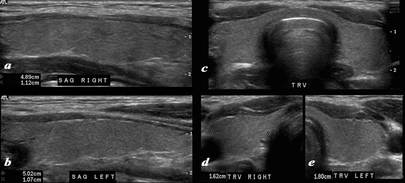
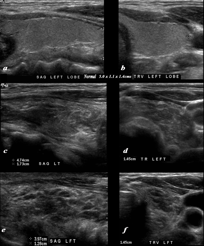
The Normal Ultrasound |
|
A normal ultrasound of the thyroid gland is demonstrated. Image a and b are sagittal images, image c is a transverse view of both lobes and the isthmus, and image d and e are individual measurements of the right and left lobe respectively.. The normal right lobe (a,d) measures 4.9cms (length) x 1.1cms (A-P anteroposterior) X 1.6 cms (transverse TRV) and the left lobe (b,e) 25.09cms (craniocaudad), by 1.1cms (A-P) by 1.8cms (transverse). Courtesy Ashley Davidoff MD Copyright 2010 93832cL.8 |
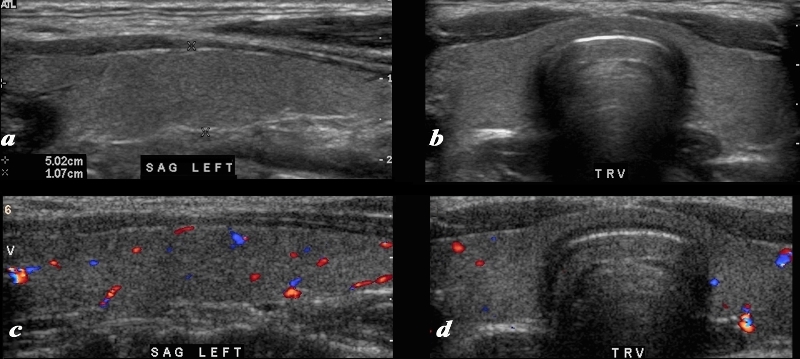
The Normal Gland and Doppler Flow |
|
A normal ultrasound of the thyroid gland is demonstrated. Image a and b are sagittal and transverse images, and images c and d are comparable views with color flow Doppler. The normal ultrasonographic characteristics demonstrating size shape position and character of the thyroid and normal color flow pattern is well demonstrated. Courtesy Ashley Davidoff MD Copyright 2010 93832c02L.8 |
Diffuse Abnormality
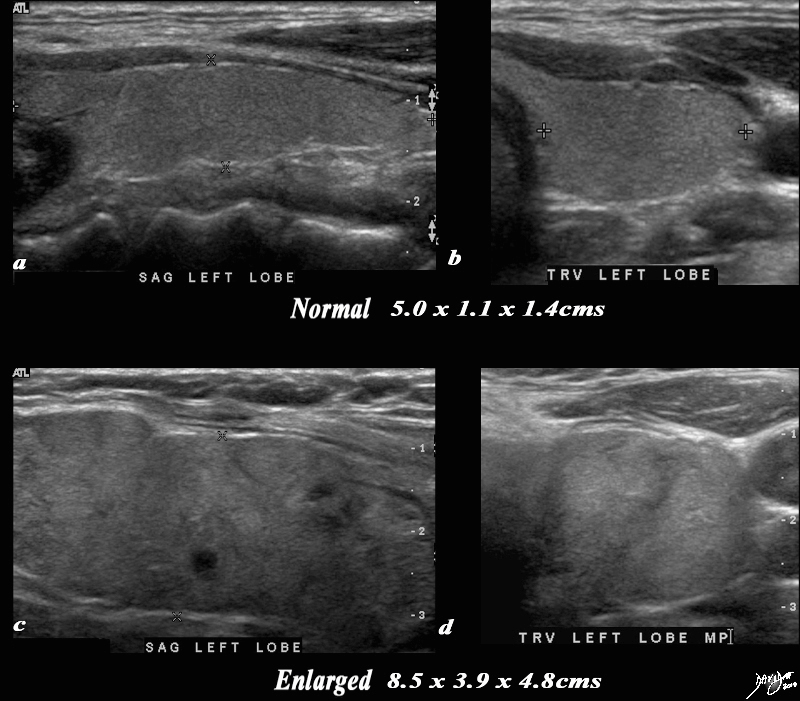
Measuring Size, Evaluating Shape and Character |
|
A normal ultrasound of the thyroid gland (a,b) is juxtaposed with the ultrasound of a 78 year female with hypothyroidism (c,d) A diffusely enlarged, heterogeneous thyroid gland is seen in the hypothyroid patient. The normal thyroid measures 5.0cms (length) x 1.1cms (A-P anteroposterior) X 1.4 cms (transverse TRV) and the abnormal thyroid measures 8.5cms (craniocaudad), by 3.9cms (A-P) by 4.8cms (transverse). Clinical findings were consistent with thyroiditis, with biochemical findings suggesting hypothyroidism. The enlarged gland in the transverse dimension is almost round. (d). This rounded shape in the transverse dimension is a clue to the presence of the enlarged gland, even before the measurements are taken and evaluated. The gland is heterogeneous particularly well seen on the sagittal view (c). The findings are consistent with a thyroiditis. Courtesy Ashley Davidoff MD Copyright 2010 94549c05g03.8s |
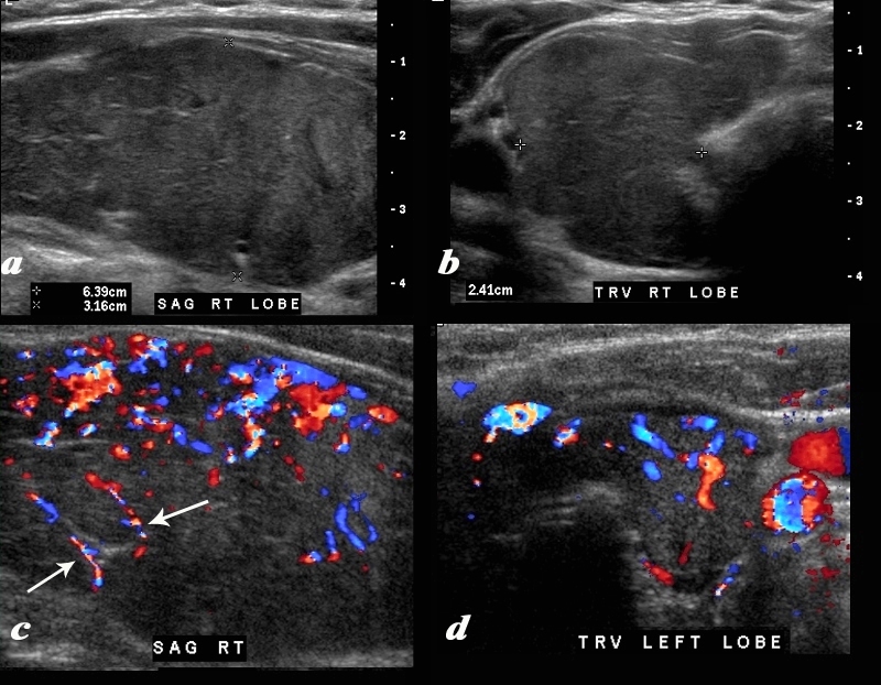
Hypervascularity of Thyroiditis |
|
A diffusely enlarged, heterogeneous thyroid gland is seen in this 30 year old hypothyroid female patient. The thyroid measures 6.4cms (craniocaudad), by 3.2cms (A-P) by 2.4cms (transverse). Clinical findings were consistent with thyroiditis, with biochemical findings suggesting hypothyroidism. The sagittal view shows coarse heterogeneous echo texture with fine white bands consistent with fibrosis. The increased vascularity is seen throughout the gland (c,d), but is also seen particularly along some of the bands in the posterior aspect of the gland (c arrows). The enlarged gland in the transverse dimension is almost round. (b). This rounded shape in the transverse dimension is a clue to the presence of the enlarged gland, even before the measurements are taken and evaluated. The findings are consistent with a thyroiditis. Courtesy Ashley Davidoff MD Copyright 2010 94583c01.8 |
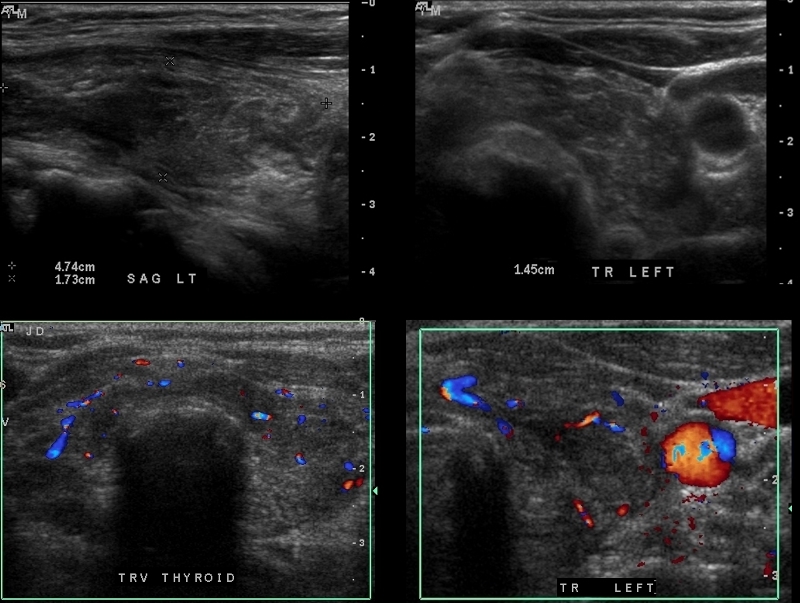
Lymphoma of the Thyroid Normal Vascularity |
|
The patient is a 62 year female who had a biopsy proven lymphoma of the thyroid. A heterogeneous thyroid gland is seen in this 62 year old female patient. The left lobe of the thyroid measures 4.7cms (craniocaudad), by 1.7cms (A-P) by 1.5cms (transverse) which is normal in size. The sagittal view shows coarse heterogeneous echo texture. The transverse view shows a rounded appearance and although the dimensions are normal the shape suggests abnormality. Additionally the isthmus is thicker than usual. The color flow Doppler study also is normal The differential diagnosis for this appearance includes thyroiditis and other infiltrative diseases such as amyloidosis. Biopsy showed lymphoma. Courtesy Ashley Davidoff MD Copyright 2010 95824c.8 |
Normal (a,b) Lymphoma (c,d) and Post Chemo XRT (d,e) |
|
The series ultrasounds demonstrate a normal patient (a sagittal ,b transverse) showing normal texture juxtaposed studies in the same projections of a 62 year female who had a biopsy proven lymphoma ((c,d). Following chemotherapy and radiation therapy she became hypothyroid and repeat ultrasound of her thyroid gland (e,f) are shown. The second series (c,d) with active lymphoma show a rounded gland in the transverse view, a thickened isthmus, with heterogeneous texture. The linear dimensions of the gland are within normal limits Following therapy the gland overall remains the same size but the texture shows many more hypoechoic amorphous regions likely due to necrosis. Courtesy Ashley Davidoff MD Copyright 2010 95824c01b.8sL |
Nodules
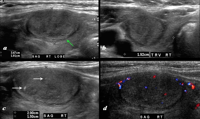
Malignant Nodule Occupying the Entire Gland |
|
A large nodule in the thyroid occupies almost the entire right lobe (a). The nodule measures 2.9cms by 1.5cms. The gland is not enlarged and measures 3.5cms (craniocaudad), by 1.8cms (A-P) by 2.4cms (transverse). The nodule is almost isoechoic with normal thyroid but shows internal irregular areas of hypoechogenicity, regions of isoechogenicity, as well as microcalcifications (white arrows (c). There is irregularity of the border at the posterior aspect of the nodule green arrow a). The halo shows irregular borders in this region as well. Internal vascularity is minimal (d). The irregular surface is concerning for a malignant processes. The diagnosis in this patient was papillary carcinoma Courtesy Ashley Davidoff MD Copyright 2010 74909c02L.8 |