The Common vein Copyright 2010
Types of Microfollicular Lesions
The finding of a microfollicular histology on a fine needle aspirate raises concern and warrants surgical resection.
|
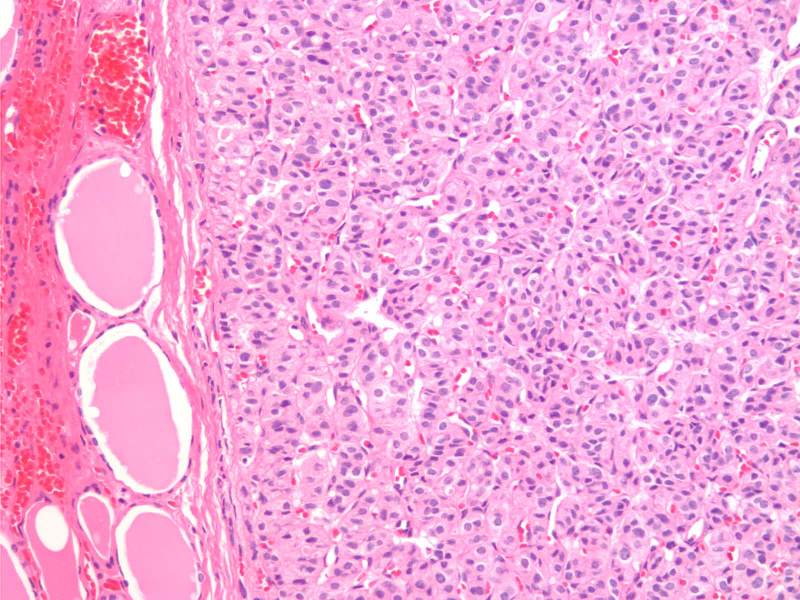
Microfollicular Adenoma H&E 20X |
|
The histological section at 20X magnification using H&E stain shows a follicular adenoma with microfollicles. The microfollicles appear as glandular structures with well defined lumens that are either empty. The size of the follicles are very similar. To the left of the image you can see the collagen capsule, and normal follicles outside the capsule. Image Courtesy Ashraf Khan MD. Department of Pathology, University of Massachusetts Medical School. 99396.8 |
|
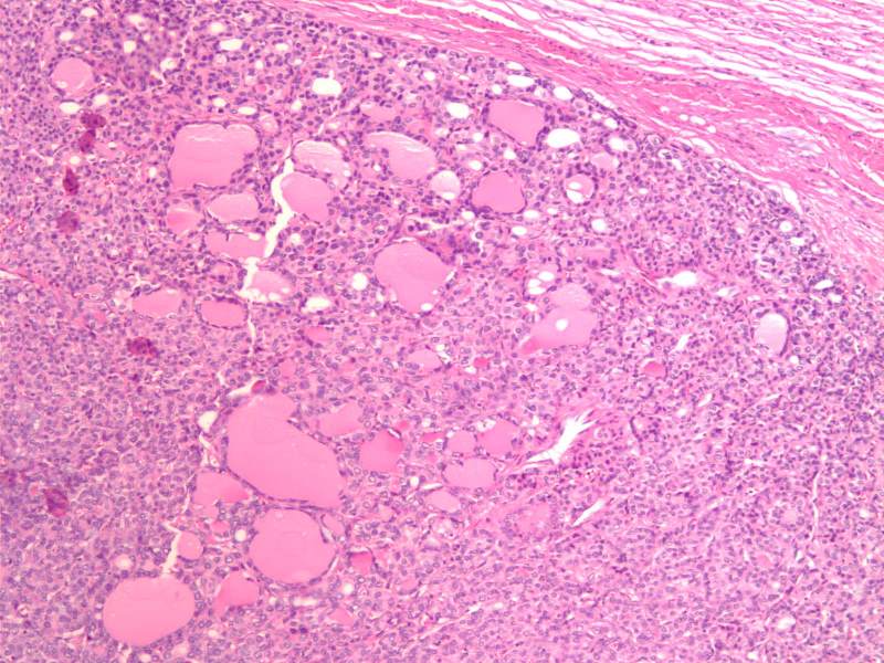
Encapsulated Follicular Variant of Papillary Carcinoma 10X H&E |
|
The histological section at 10X magnification using H&E stain shows a encapsulated follicular variant of papillary carcinoma (FVPC) with microfollicular architecture. Image Courtesy Ashraf Khan MD. Department of Pathology, University of Massachusetts Medical School. 99397.8 |
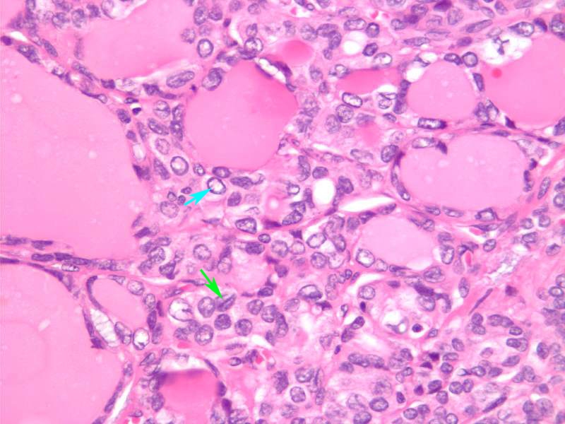
Encapsulated Follicular Variant of Papillary Carcinoma 40X H&E |
|
The histological section at 40X magnification using H&E stain shows a encapsulated follicular variant of papillary carcinoma (FVPC) with microfollicular architecture. This high power view shows papillary cancer nuclei with Orphan Annie nuclei and grooves. Orphan Annie nuclei a “cleared-out” or empty appearance, similar to Little Orphan Annie’s eyes (light blue arrow). Nuclear grooves characteristic lines that run across the nuclei (green arrow). Image Courtesy Ashraf Khan MD. Department of Pathology, University of Massachusetts Medical School. 99398.81 |
|
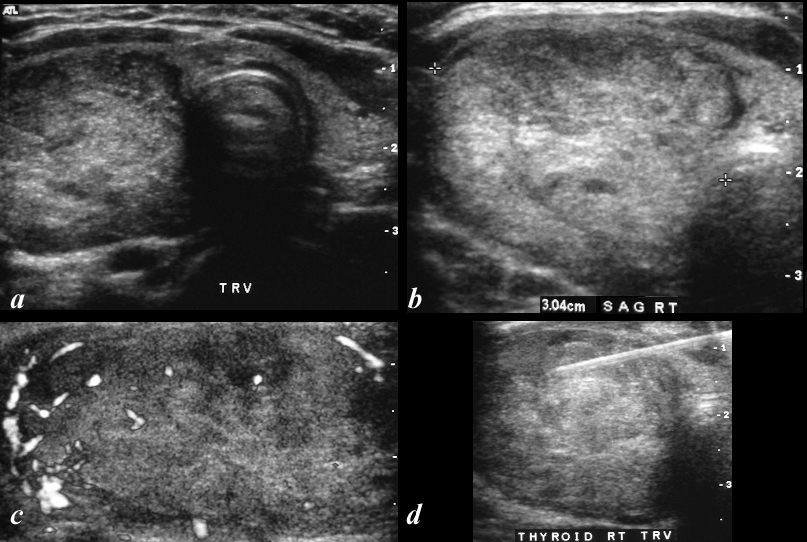
Microfollicular Lesion – Microfollicular Adenoma |
|
The ultrasound is from an adult female and shows a 3cms mostly iso to hyperechoic lesion (a,b) in the right lobe of the thyroid occupying almost the entire lobe. No obvious calcifications nor halo is present and the vascularity is mostly peripheral (c) A biopsy (d) supported the diagnosis of a microfollicular lesion. In the presence of microfollicles – surgical resection is indicated. Courtesy Barry Sacks MD 96970c.8L |