
The Common Vein Copyright 2010
Introduction
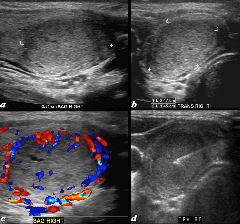
Hyperechoic Nodule
Hurthle Cell Neoplasm |
|
This ultrasound is from a female patient who presents with a thyroid nodule. Ultrasound shows a mass in the right lobe of the thyroid. The nodule measures 2.5cms in sagittal (a), 2.2 cms in A-P dimension (b) and 1.5cms in transverse view. The mass is isoechoic to hyperechoic, is mostly homogeneous, but for tiny cystic spaces with Doppler flow that is mostly peripheral but does have some central flow as well (c). Image (d) shows a well placed aspiration needle. A diagnosis of Hurthle cell neoplasm was made. Courtesy Barry Sacks MD Copyright 2010 97073cL.8 |