
The Common vein Copyright 2010
Introduction
The thyroid is surrounded by a rich network of lymphatics. There are approximately 300 lymph nodes in the neck, and flow is across the midline and also superiorly and inferiorly.
The most immediate lymph nodes include the prelaryngeal lymph node of delphian, the pretracheal lymph nodes and the paratracheal lymph nodes. These nodes are close the the recurrent laryngeal nerve.
Two basic Systems are present
Superficial System
Nodes that lie superficial to the sterno cleidomastoid
Deep System
Related to the Internal Jugular Vein
upper
middle
lower
Lymphatic Drainage – Anterior View |
|
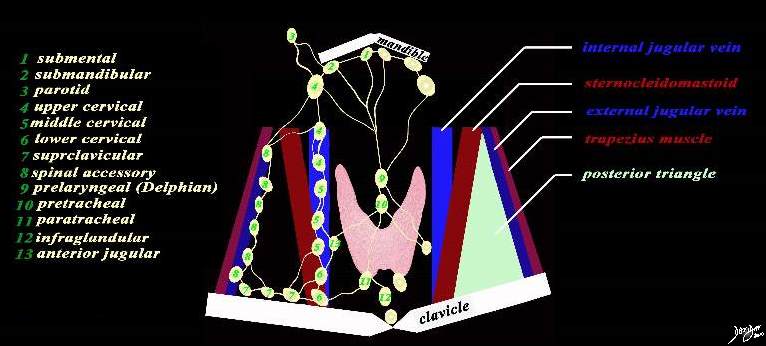
There are about 13 named sets of lymph nodes that serve to drain the neck and the region of the thyroid gland. The submental lymph nodes (1) lie inferior to the anterior belly of the digastric muscle. The submandibular glands (2) lie under the mandible and between the two lims of the digastric muscle. The parotid lymph nodes (3) lie anterior to the ear The cervical lymph nodes are in close association of the internal jugular vein and sternocleidomastoid muscle. Their division of upper middle and lower nodes relates to bony landmarks. The upper cervical nodes (4) lie above the hyoid bone, the middle group (5) lie between the hyoid bone and cricoid cartilage, while the lower group (6) lie between the cricoid and clavicle. The supraclavicular group (7) lie above the clavicle, and the posterior traingle group (accessory nodes) lie high in th posterior triangle between the sternocleidomastoid and trapezius. The nodes that relate to the posterior triangle and external jugular vein are the spinal accessory nodes (aka posterior triangle nodes) (8). Above the thyroid and in the midline is the prelaryngeal node (Delphian node 9), and just below it and also in the midline is the pretracheal node (10) A pair od p[aratracheal nodes lie below the thyroid (11), and a midline infraglandular node also lies below thr gland a sits name implies. The anterior jugular vein has a node associate with it and its is called the anterior jugular node (13) The posterior triangle (light green) is formed by the posterior border of the sternocleidomastoid, the anterior border of the trapezius, and the base is formed by the clavicle. Courtesy Ashley Davidoff MD copyright 2010 all rights reserved 94885b06b04L03.8s |
Conceptual Framework of the Cervical Lymph Nodes |
|
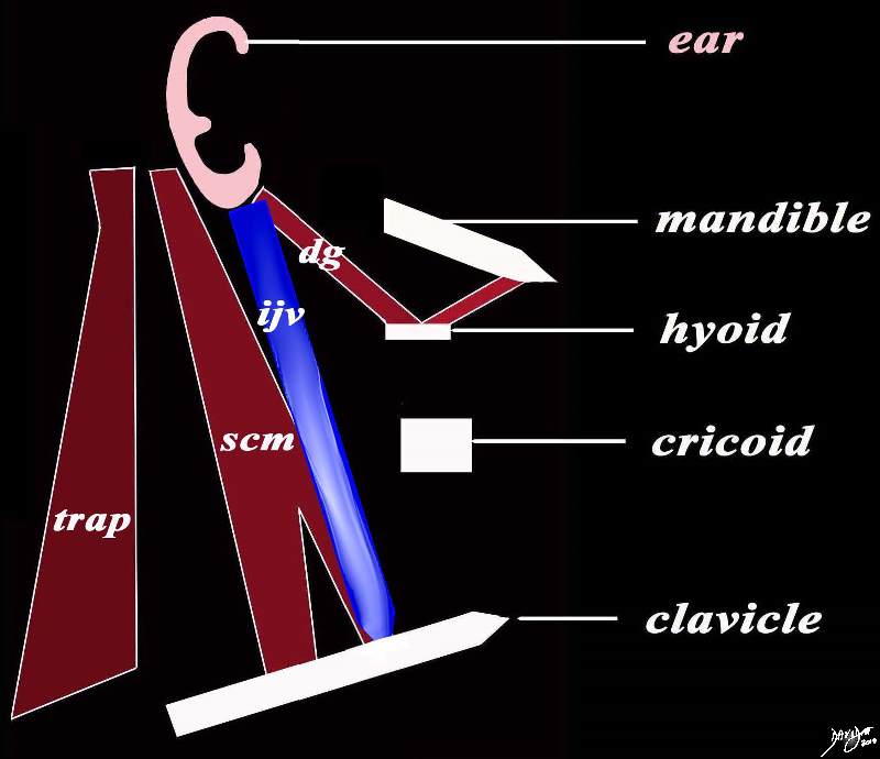
The cervical lymph nodes have muscular and bony/cartligenous landmarks extending from the ear superiorly to the clavicle inferiorly. The bony landmarks starting superiorly include the inferior margin of the mandible, the hyoid bone, the cricoid cartilage and the clavicle. The muscles include the digastric muscle (dg) with anterior and posterior bellies superiorly, then the sternocleidomastoid (scm), and finally the trapezius (trap). The internal jugular vein (blue) is a central landmark for a large number o the cervical nodes. Courtesy Ashley Davidoff MD copyright 2010 all rights reserved 94884b06b03.8s |
The Normal Distribution of 300 Lymph Nodes of the Neck |
|
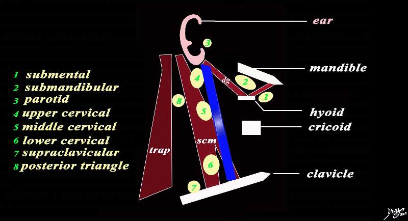
There are 8 basic sets of lymph nodes relating to the structures described. The submental lymph nodes (1) lie inferior to the anterior belly of the digastric muscle. The submandibular glands (2) lie under the mandible and between the two lims of the digastric muscle. The parotid lymph nodes (3) lie anterior to the ear The cervical lymph nodes are in close association of the internal jugular vein and sternocleidomastoid muscle.. Their division of upper middle and lower nodes relates to bony landmarks. The upper cervical nodes (4) lie above the hyoid bone, the middle group (5) lie between the hyoid bone and cricoid cartilage, while the lower group (6) lie between the cricoid and clavicle. The supraclavicular group (7) lie above the clavicle, and the posterior traingle group (accessory nodes) lie high in th posterior triangle between the sternocleidomastoid and trapezius. Courtesy Ashley Davidoff MD copyright 2010 all rights reserved 94884b11.8s |
Metatstattic Calcified Nodes of Papillary Thyroid Carcinoma At the level of the hyoid bone |
|
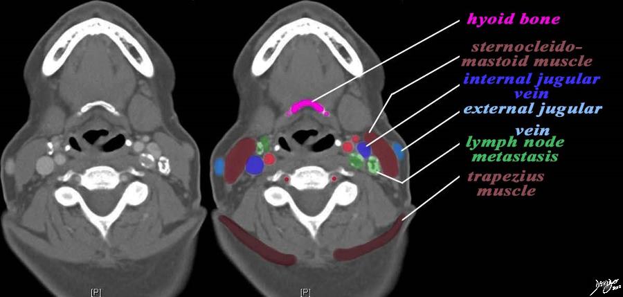
The CT scan is from a middle aged female patient with metastatic papillary carcinoma of the thyroid. her nodes contain calcification and hence are easily seen. The case exemplifies the metastatic lymph node pattern of thyroid carcinoma. The most superior cut is at the level of the hyoid bone (pink). At this level the nodes associated with the internal jugular vein (royal blue) are the upper cervical nodes. These are posterior to the internal vein on the left and anterior on the left (dark green) There are no nodes associated with the external jugular vein nor in the posterior triangle which lies between the sternocleidomastoid muscle anteriorly and the trapezius posteriorly (maroon). Courtesy Ashley Davidoff MD copyright 2010 all rights reserved 93812c14.9s |
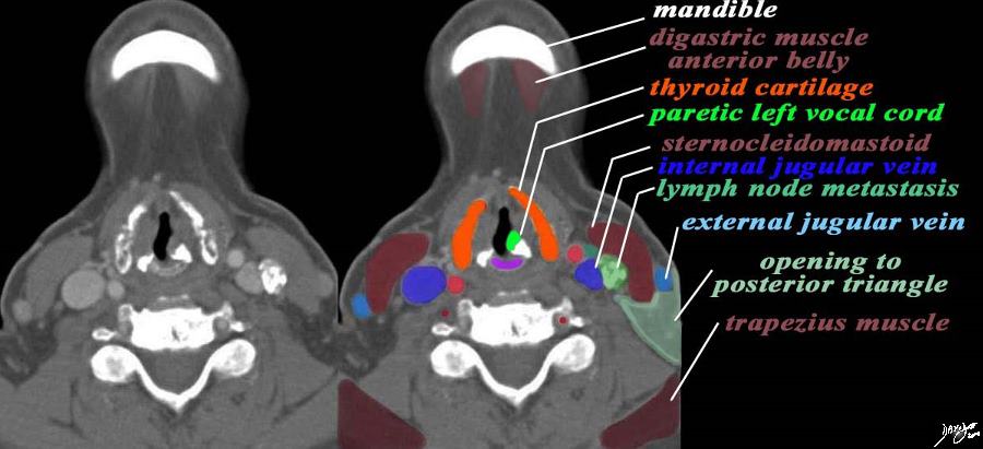
At the Level of the Thyroid Cartilage |
|
The CT scan is from a middle aged female patient with metastatic papillary carcinoma of the thyroid. Her nodes contain calcification as seen in the image on the left and hence are easily seen. The case exemplifies the metastatic lymph node pattern of thyroid carcinoma. This cut is at the level of the tyhroid cartilage (orange) and the the most superior aspect of the cricoid cartilage (prurple) . At this level the nodes associated with the internal jugular vein (royal blue) are the middle cervical nodes (green nodes). These lie lateral to the internal jugular vein (royal blue) on the left . There is an adducted left vocal cord (bright green) suggesting that it is paretic. There is a region of exudation or a small nodule (dark green) abutting the left carotid artery (bright red), The recurrent laryngeal nerve is intimately associated with the inferior thyroidal artery There are no nodes associated with the external jugular vein nor in the posterior triangle (light green triangular patch) which lies between the sternocleidomastoid muscle anteriorly and the trapezius posteriorly (maroon). Courtesy Ashley Davidoff MD copyright 2010 all rights reserved 93812c16b.9s |
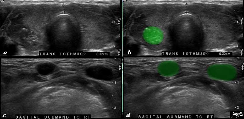
Submental Lymph Nodes Papillary Carcinoma |
|
The thyroid ultrasound is from a 31 year old female with papillary carcinoma of the right lobe of the thyroid (b, bright green). Two smallsubcentimeter lymph nodes are identified in the submental area lymph nodes They are almost anechoic Courtesy Ashley DAvidoff MD copyright 2010 all rights reserved 94920c02.8s |
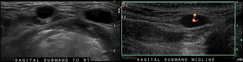
Submental Lymph Nodes with Central Vascularity |
|
The thyroid ultrasound is from a 31 year old female with papillary carcinoma of the right lobe of the thyroid. Two smallsubcentimeter lymph nodes are identified in the submental area. They are almost anechoic but demonstrate central vascularity. Courtesy Ashley Davidoff MD copyright 2010 all rights reserved 94920c01b.8s |
References
Reede, DL, Bergeron, RT, Whelan, M A , Cohen, NL, Persky, MS Computed Tomography of Cervical Lymph Nodes RadioGraphics 339 Volume 3, Number 2 June 1983
Silverman P Cancer Imaging Lymph Node Imaging in Cancer Imaging. 2005; 5(Spec No A): S57–S67. Published online 2005 November 23 5star
Ying MTC Ahuja AT Ultrasonographyof the Cervical Lymph Nodes CUHK.edu.hk 5star