
The Common vein Copyright 2010
Definition
Lymphoma of the thyroid is a primary malignant condirion that arises in the thyroid gland. Thyroid lymphoma is frequently non-Hodgkins type and is often associated with Hashimoto’s thyroiditis.
Thyroid Lymphoma is thought to be caused by chronic antigen stimulation (such as that in hashimoto’s) that leads to proliferation of lymphoid tissue in the thyroid gland. The chronic stimulation and proliferation can eventually lead to genetic mutations resulting in a malignant lymphoma.
Structural changes include a rapidly enlarging firm thyroid mass. Normal thyroid gland tissue is replaced with malignant lymphocytes. There may not be any functional changes, but 30-40% of patients with primary thyroid lymphoma will have hypothyroidism, either secondary to loss of thyroid tissue or underlying Hashimoto’s disease.
Thyroid lymphoma presents clinically as a rapidly enlarging thyroid mass, often with concurrent cervical lymphadenopathy. Often, patients will have underlying Hashimoto’s thyroiditis and symptoms of hypothyroidism. Patients with more extensive disease may present with hoarseness, dysphagia, and/or cough.
Imaging of thyroid lymphoma is done with thyroid ultrasound and CT scan. Ultrasound can show size and characteristics of thyroid mass, allows for evaluation of lymph node involvement, and guides biopsy. CT scan can assess for local invasion and metastatic disease. Diagnosis is best made with FNAB or excisional biopsy.
Treatment for thyroid lymphoma is similar to treatment of other lymphomas and depends on the lymphoma type and staging. Typical treatment regimens involve chemotherapy, specifically CHOP, followed by local radiation therapy.
Enlarged Heterogeneous Gland |
|
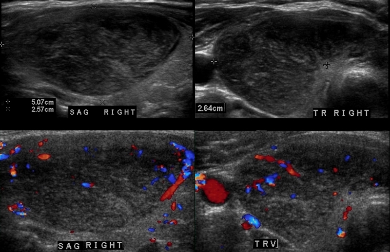
A large mostly isoechoic enlarged thyroid is demonstrated on the ultrasound of this 64 year female. Images a and b are in the sagittal projection and b and d in the transverse plane. The lobes of the thyroid measure 5.1cms in craniocaudad span by 2.6 cms in A-P dimension by 2.6cms transversely. Note the rounded shape of the right lobe suggesting enlargement of the gland by virtue of shape. The isthmus is also thickened and measured almost a cms. The left lobe had similar size shape and echotexture. The parenchyma is heterogeneous with no nodules (a,b) and Doppler flow is normal (c,d) and unremarkable. A biopsy showed B cell lymphoma Courtesy Ashley Davidoff MD Copyright 2010 94882c.8 |
Lymphoma – a Second Case |
|
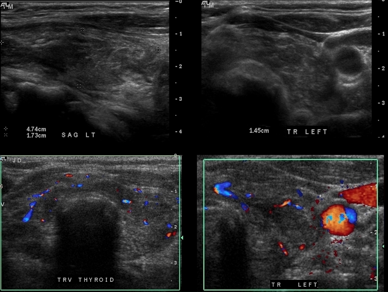
The patient is a 62 year female who had a biopsy proven lymphoma of the thyroid. A heterogeneous thyroid gland is seen in this 62 year old female patient. The left lobe of the thyroid measures 4.7cms (craniocaudad), by 1.7cms (A-P) by 1.5cms (transverse) which is normal in size. The sagittal view shows coarse heterogeneous echo texture. The transverse view shows a rounded appearance and although the dimensions are normal the shape suggests abnormality. Additionally the isthmus is thicker than usual. The color flow Doppler study also is normal The differential diagnosis for this appearance includes thyroiditis and other infiltrative diseases such as amyloidosis. Biopsy showed lymphoma. Courtesy Ashley Davidoff MD Copyright 2010 95824c.8 |
Lymphoma of the Thyroid Same Patient as Above |
|
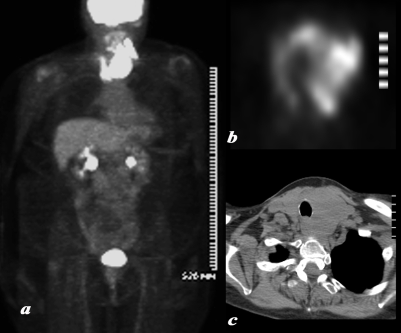
A large and very PET avid left lobe of the thyroid is demonstrated on the body PET scan shown in full body view in (a) and in transverse view of the thyroid in (b). The patient is a 62 year female who had a biopsy proven lymphoma of the thyroid.. The CT shows asymmetric and smooth and homogeneous enlargement of the thyroid. The findings are consistent with the final biopsy proven diagnosis of lymphoma. Courtesy Ashley Davidoff MD Copyright 2010 95855c01.8 |

PET following Chemotherapy and Radiation Therapy |
|
The patient is a 62 year female who had a biopsy proven lymphoma of the thyroid . A large and very PET avid left lobe of the thyroid is demonstrated on the body PET scan shown in full body view in (a) with resolution of the mass and significant and likely remission changes (b) following chemotherapy and radiotherapy. At the base of the neck there is a U shaped pattern corresponding to residual thyroid tissue. The SUV was 5, higher than typically expected which usually ranges between 1and 2 suggesting persistent moderate uptake. This is likely due to radiation thyroiditis. The findings are most consistent with lymphoma in remission following therapy and likely associated XRt thyroiditis. Courtesy Ashley Davidoff MD Copyright 2010 95855c02.8 |
Normal (a,b), Lymphoma (c,d) and Lymphoma post Chemotherapy and XRT (e,f) Same Patient as Above |
|
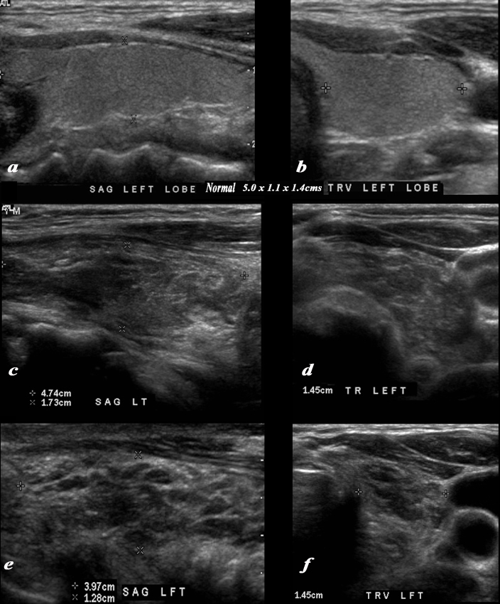
The series ultrasounds demonstrate a normal patient (a sagittal ,b transverse) showing normal texture juxtaposed studies in the same projections of a 62 year female who had a biopsy proven lymphoma ((c,d). Following chemotherapy and radiation therapy she became hypothyroid and repeat ultrasound of her thyroid gland (e,f) are shown. The second series (c,d) with active lymphoma show a rounded gland in the transverse view, a thickened isthmus, with heterogeneous texture. The linear dimensions of the gland are within normal limits Following therapy the gland overall remains the same size but the texture shows many more hypoechoic amorphous regions likely due to necrosis. code thyroid shape size character homogeneous heterogeneous Courtesy Ashley Davidoff MD Copyright 2010 95824c01b.8sL |