
The Common Vein Copyright 2010
Introduction
The thyroid loses volume with aging and in fact metabolism gradually slows beginning around age 20. However functionally aging only affects the thyroid in minor ways with a slight decrease in T3 levels. Disease of the thyroid such as autoimmnune disorders become more prevalent with age.
The incidence of thyroid nodules increases with age.
50% of 50 year olds will have at least one thyroid nodule.
60% of 60 year olds will have at least one thyroid nodule.
70% of 70 year olds will have at least one thyroid nodule.
|
Normal and Atrophied Gland |
|
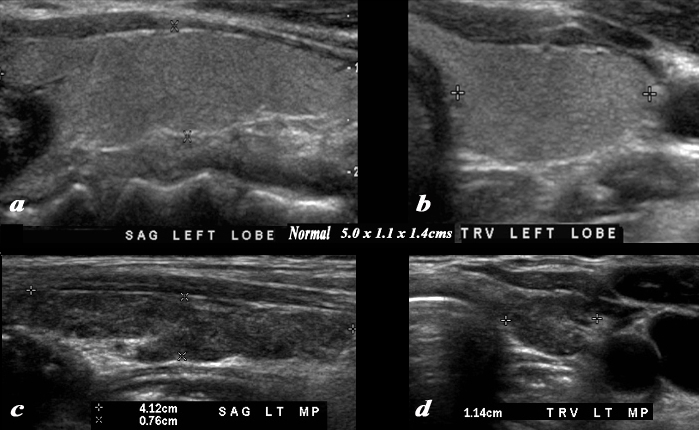
The patient is a 70year female who shows diffuse atrophy of her thyroid by this ultrasound examination. A normal thyroid gland in sagittal (a) and transverse (b) planes is juxtaposed on the atrophied gland(c,d) The gland is mildly heterogeneous. The left lobe of the thyroid measures 4.1cms (craniocaudad), by .8 cms (A-P) by 1.1cms (transverse). The sagittal view shows mildly coarse heterogeneous echo texture. The transverse view shows a normal shape. The isthmus is normal in size. These findings suggest age related involution of the gland. Courtesy Ashley Davidoff MD Copyright 2010 94503c02L |
|
Age Related Involution of the Thyroid |
|
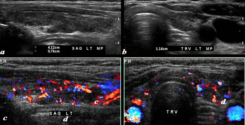
The patient is a 70 year female who shows diffuse atrophy of her thyroid by this ultrasound examination. The gland is mildly heterogeneous. The left lobe of the thyroid measures 4.1cms (craniocaudad), by .8 cms (A-P) by 1.1cms (transverse). The sagittal view shows mildly coarse heterogeneous echo texture. The transverse view shows a normal shape. The isthmus is normal in size. The color flow Doppler study seems hypervascular but the significance of this finding is not known. These findings suggest age related involution of the gland. Courtesy Ashley Davidoff MD Copyright 2010 94503c.8L |
CT Scan
Ther density of the thyroid on CT scan decreases with age.
97362c01L01 |
|
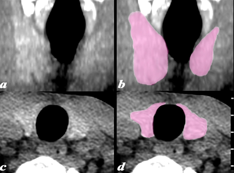
This normal CTscan is from a 28year old normal male and reveals the slight increase in density of the thyroid compared to normal surrounding soft tissue. This is due to the normal uptake of iodine. Image a and b show a reconstructed coronal view and images c and d are in the axial plane Courtesy Ashley Davidoff MD Copyright 2010 97362c01L01 |