Copyright 2011
Histology
Histological Divisions
At the histological level the unit of the thyroid is the lobule. Each lobule consists of 20-30 follicles.
The thyroid is brownish-red in color, is a highly vascular gland and is composed of multiple lobules separated by thin septations. Each lobule consists of 20-40 follicles and supplied by an intralobular artery.
The thyroid is characterized by three main histological features: colloid containing follicles, follicular epithelium, and parafollicular cells.
The follicles secrete thyroxin and triiodothyronine. The follicles are spherical cavities which contain colloid, a viscous secretion rich gel primarily a store for thyroglobulin which is the precursor for T3 and T4. The colloid serves as a reservoir for raw materials and also as storage for the hormones themselves. The follicles range in size from 50-500microns but average about 200 microns in diameter. There is sufficient storage of thyroid hormone for about 100days.
|
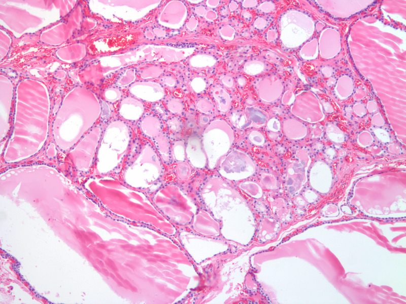
Follicles – Normofollicular and Macrofollicular 10X H& E |
|
The histological section at 10X magnification using H&E stain shows normal thyroid tissue with normofollicular and macrofollicular changes. The single layer of epithelium surrounding the follicle is made up of a simple cuboidal cells and the lumen is filled with pink staining colloid. Thyroglobulin and iodide are contained within the colloid and are used to produce thyroid hormones including thyroxine. Between the cells are the parafollicular cells that produce calcitonin. Image Courtesy Ashraf Khan MD Department of Pathology, University of Massachusetts Medical School. 99411.8 |
Each follicle is lined by a single layer of follicular epithelial cells, collectively termed the follicular epithelium, whose morphology ranges from cuboid when inactive to columnar when active. These cells trap iodine from the bloodstream and use it to synthesize thyroxine and triiodothyronine from the glycoprotein thyroglobulin. If not needed immediately these hormones remain stored in thyroglobulin and are secreted along with colloid into the follicles. When serum levels of thyroxine and triiodothyronine drop too low colloid is absorbed back into the thyroid cells where thyroglobulin is cleaved and the hormones are released through the base of the cell into the surrounding capillaries.
Parafollicular cells or C cells surround the follicular epithelium and follicles and synthesize calcitonin, which help regulate calcium metabolism in conjunction with the parathyroid glands.
|
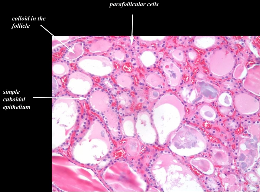
Transitional Epithelium Follicles, Colloid, and Parafollicular Cells 20X H&E |
|
The histological section at 20X magnification using H&E stain shows normal thyroid tissue with normofollicular and macrofollicular changes. The single layer of epithelium surrounding the follicle is made up of a simple cuboidal cells and the lumen is filled with pink staining colloid. Thyroglobulin and iodide are contained within the colloid and are used to produce thyroid hormones including thyroxine. Between the cells are the parafollicular cells that produce calcitonin. Image Courtesy Ashraf Khan MD. Department of Pathology, University of Massachusetts Medical School. 99412.8b02 |
Normofollicles and Macrofollicles
|
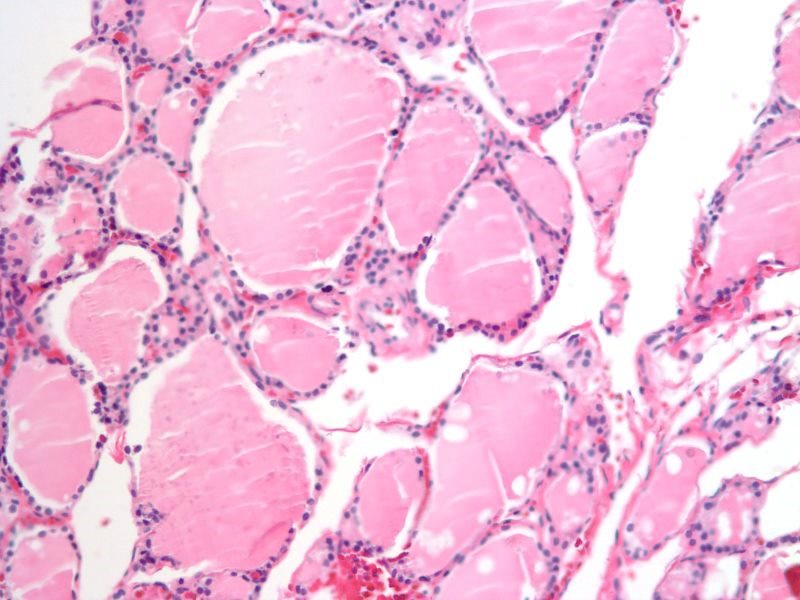
Normal Follicular Appearance Normofollicles and Macrofollicles 20X H&E |
|
The histological section at 20X magnification using H&E stain shows normal thyroid tissue with normofollicular and macrofollicular changes. The single layer of epithelium surrounding the follicle is made up of a simple cuboidal cells and the lumen is filled with pink staining colloid. The different size of the follicles is normal and has no pathological implications. Thyroglobulin and iodide are contained within the colloid and are used to produce thyroid hormones including thyroxine. Between the cells are the parafollicular cells that produce calcitonin. Image Courtesy Ashraf Khan MD. Department of Pathology, University of Massachusetts Medical School. 99410.81 |
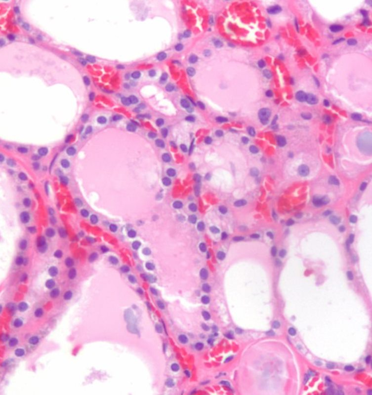
Epithelium and Capillary Network
|
Epithelium and Capillary Network |
|
The histological section using H&E stain shows normal thyroid tissue with a focus on a bilobed follicle toward left of centre. The cuboidal epithelium is exemplified in this follicle. An extensive capillary network in close association with the glandular tissue is typical of an endocrine organ. The capillaries are identified by the red cells lined up within the vessel lumen. Image Courtesy Ashraf Khan MD. Department of Pathology, University of Massachusetts Medical School. 99412d |
|
The Thyroid Gland – Unique in the Body |
|
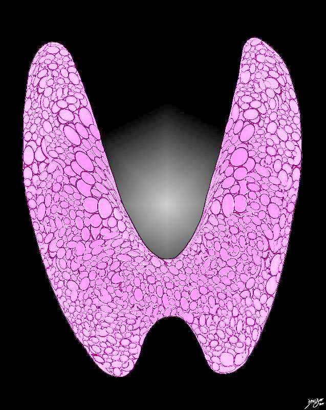
Artistic rendition of the normal thyroid gland The histological make up of the thyroid has a unique appearance consisting of variably sized rounded glandular rosettes with follicles that contain a homogeneously pink material called colloid, subtended by a basal layer of a single layer of cuboidal cells that secrete and extract material from the colloid when needed. Courtesy Ashley Davidoff MD copyright 2011 all rights reserved 94460b13t06p03.81s |