
The Common Vein Copyright 2010
Introduction
Simple
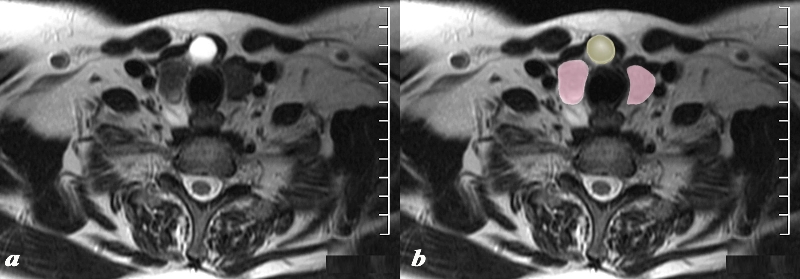
Thyroglossal Duct Cyst – MRI T2 Weighted Sequence |
|
The MRI T2 weighted image through the inferior aspect of the thyroid gland (pink)shows a cystic structure (yellow), intensely T2 bright, in the region of the isthmus in the ventral aspect of the gland. Findings are consistent with a thyroglossal cyst Courtesy Ashley Davidoff MD Copyright 2010 97310cL.8 |
Complex
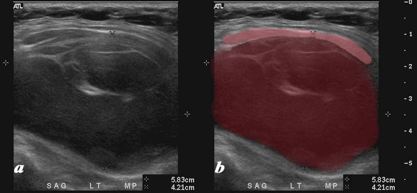
Hemorrhagic Cyst Presenting as a Goiter |
|
A large mostly hypoechoic mass expands the left lobe of the thyroid on the ultrasound of this 54 year female who presents with an enlarged thyroid gland. The left lobe is filled with low level echoes and contains multiple thin septal echogenic walls (maroon). The mass appears to displace and compress normal soft tissues of the neck (pink) anteriorly. Doppler showed no flow. The mass occupies the entire left lobe and measures 5.8cms in craniocaudad span and by 4.2 cms transversely The diagnosis in this patient is a hemorrhagic cyst causing a goiter. Courtesy Ashley Davidoff MD Copyright 2010 94656c01L |