
The Common vein Copyright 2010
Introduction
What does it look and feel like?
The thyroid is a brownish-red in color, soft to the feel.
The imaging characteristics of the thyroid are far more revealing. These characteristics are most easily observed using ultrasound.
The Normal Ultrasound
|
Homogeneous Echotexture of the Thyroid |
|
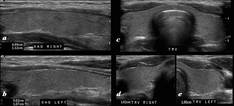
A normal ultrasound of the thyroid gland is demonstrated. Image a and b are sagittal images, image c is a transverse view of both lobes and the isthmus, and image d and e are individual measurements of the right and left lobe respectively.. The vascularity in the thyroid makes it slightly more echogenic than the surrounding muscle, and its ultrasound texture is reminiscent of testicular echogenicity. Courtesy Ashley Davidoff MD Copyright 2010 93832cL.8 |
|
Normal Ultrasound and Doppler Examination |
|
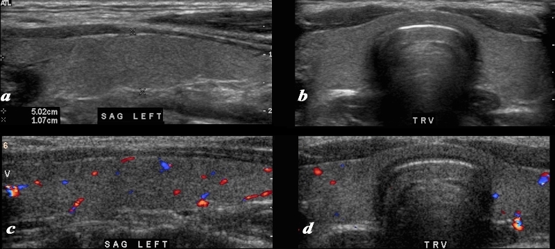
A normal ultrasound of the thyroid gland is demonstrated. Image a and b are sagittal and transverse images, and images c and d are comparable views with color flow Doppler. The normal ultrasonographic characteristics demonstrating size shape position and character of the thyroid and normal color flow pattern is well demonstrated. Note the relatively hypoechoic muscle as a thin layer just anterior to the gland and the more echogenic fat as the most superficial tissue. Courtesy Ashley Davidoff MD Copyright 2010 93832c02L.8 |
|
Normal MRI Characteristics |
|
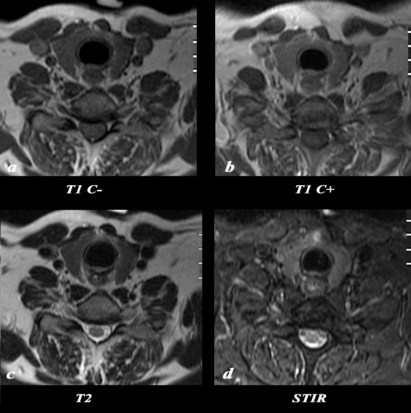
The MRI of the thyroid reveals the appearance of the thyroid on T1 weighted sequence without contrast (a), T1 with contrast (b), T2 weighted image (c), and a STIR image(d). The normal gland appears remarkably similar on all 4 phases showing mild enhancement and mild STIR hyperintensity. Courtesy Ashley Davidoff MD Copyright 2010 97332c02L.8 |
The Normal CT scan
The normal gland though small is well seen due to its slightly higher density compared to surrounding tissues as a result of its iodine content. There are differences in the density of the thyroid on CT based on geography and differences in dietary iodine uptake.
There is no difference in density between male and female though the density of the thyroid decreases with age.
|
Non Contrast CT Iodine Containing Normal Thyroid with Higher Density |
|
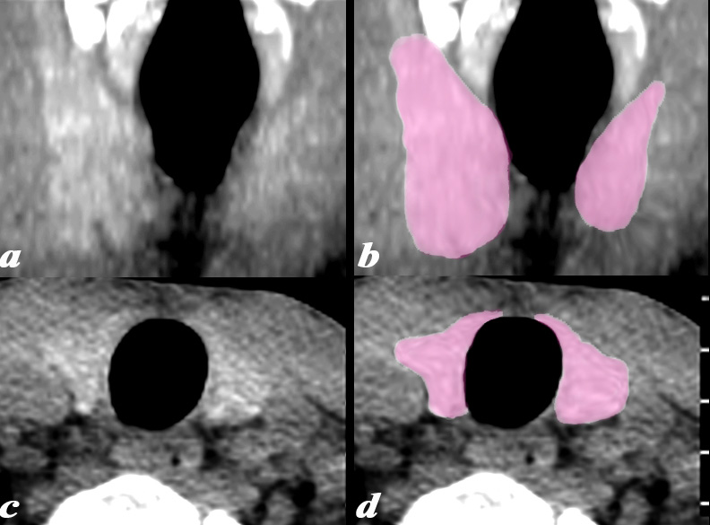
This noncontrast CTscan is from a 28year old normal male and reveals the slight increase in density of the thyroid compared to normal surrounding soft tissue. This is due to the normal uptake of iodine. Images a and b show a reconstructed coronal view and images c and d are in the axial plane Courtesy Ashley Davidoff MD Copyright 2010 97362c01L01 |
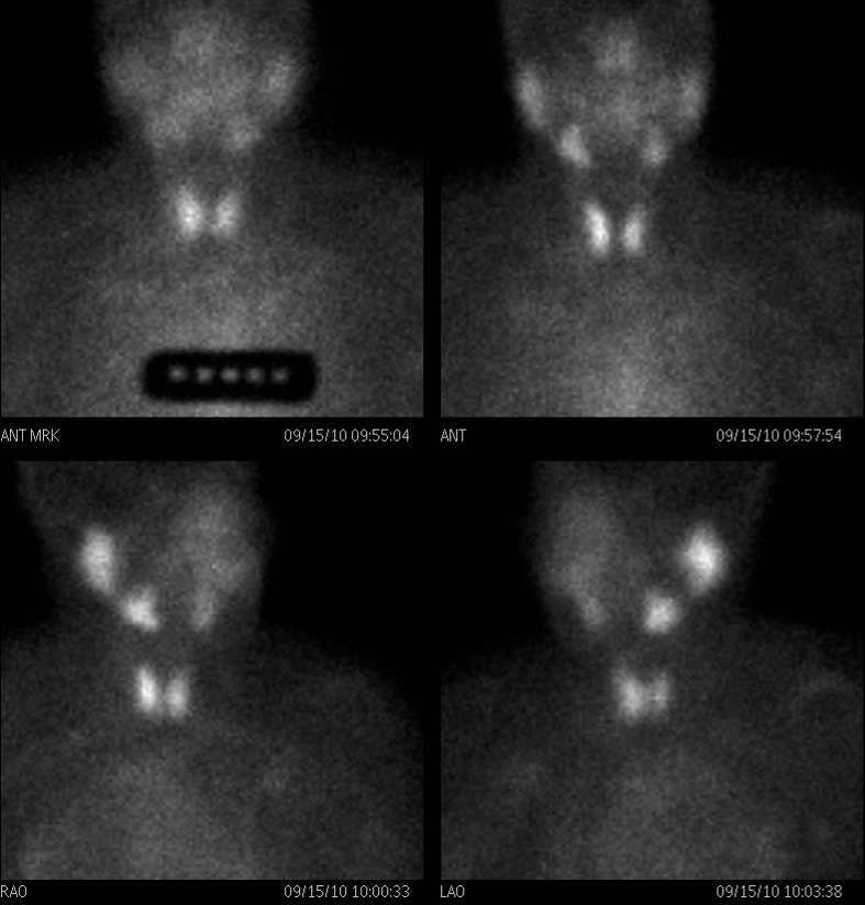
Normal Nuclear Pertechnitate Scan
|
Normal Technetium Pertechnitate Scan |
|
This is a thyroid scan from a 39 year old female with a clinical history of hyperthyroidism. She was injected with 16.1mCi of 99mTechnetium pertechnitate. The scan shows normal uniform uptake of the radioisotope in both lobes of the thyroid gland. The uptake in the thyroid gland is greater than the uptake in the submandibular and parotid glands. These findings are consistent with a normal thyroid gland. Courtesy Alan Ashare MD Copyright 2010 97511b01 |
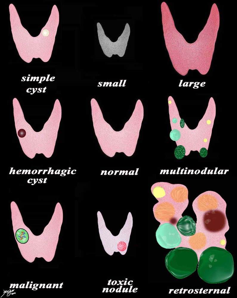
Examples of a Change in Character – Diseases
|
Changes in Character with Disease |
|
A variety of appearances of the thyroid gland in disease including (from top left to right a simple cyst (yellow) the small atrophied gland seen in hypothyroidism, the large hyperemic gland seen in acute thyroiditis and Graves disease, the hemorrhagic cyst (maroon) normal, multinodular goiter of cysts (yellow), heterogeneous (light green) and solid (dark green), the malignant nodule, hot nodule with secondary atrophy of the gland, and the large multinodular retrosternal gland. Courtesy Ashley Davidoff MD copyright 2010 all rights reserved 93852e07.9s |
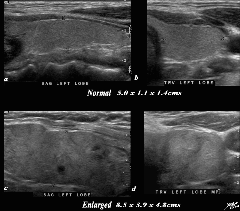
Change in Echotexture – Thyroiditis
|
Normal and the Heterogeneous Nature of Thyroiditis |
|
A normal ultrasound of the thyroid gland (a,b) is juxtaposed with the ultrasound of a 78 year female with hypothyroidism (c,d) A diffusely enlarged, heterogeneous thyroid gland is seen in the hypothyroid patient. The normal thyroid measures 5.0cms (length) x 1.1cms (A-P anteroposterior) X 1.4 cms (transverse TRV) and the abnormal thyroid measures 8.5cms (craniocaudad), by 3.9cms (A-P) by 4.8cms (transverse). Clinical findings were consistent with thyroiditis, with biochemical findings suggesting hypothyroidism. The enlarged gland in the transverse dimension is almost round. (d). This rounded shape in the transverse dimension is a clue to the presence of the enlarged gland, even before the measurements are taken and evaluated. The gland is heterogeneous particularly well seen on the sagittal view (c). The findings are consistent with a thyroiditis. Courtesy Ashley Davidoff MD Copyright 2010 94549c05g03.8s |
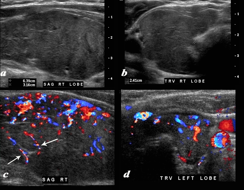
|
Thyroiditis – Enlargement Fibrous Strands and Hypervascularity |
|
A diffusely enlarged, heterogeneous thyroid gland is seen in this 30 year old hypothyroid female patient. The thyroid measures 6.4cms (craniocaudad), by 3.2cms (A-P) by 2.4cms (transverse). Clinical findings were consistent with thyroiditis, with biochemical findings suggesting hypothyroidism. The sagittal view shows coarse heterogeneous echo texture with fine white bands consistent with fibrosis. The increased vascularity is seen throughout the gland (c,d), but is also seen particularly along some of the bands in the posterior aspect of the gland (c arrows). The enlarged gland in the transverse dimension is almost round. (b). This rounded shape in the transverse dimension is a clue to the presence of the enlarged gland, even before the measurements are taken and evaluated. The findings are consistent with a thyroiditis. Courtesy Ashley Davidoff MD Copyright 2010 94583c01.8 |
Multinodular Goiter
|
Non Toxic Multinodular Gland – Normal Size |
|
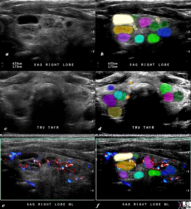
This 80 year old male presents with an asymptomatic multinodular goiter, consisting many nodules of varying size and echogenicity, as seen by sagittal ultrasound (a,b) and transverse imaging images (c,d), and Doppler imaging (e,f). The multiple nodules that are not border forming are within the confines of the parenchyma and do not alter the shape of the gland. The right lobe of the thyroid measures about 4.5 cms in sagittal 1.5cms.in A-P dimension, and 1.5cms in the transverse plane. The gland is therefore not enlarged. In addition the gland has a normal appearance in the transverse projection and the borders are not rounded to suggest enlargement. The Doppler study shows no internal vascularity in any of the nodules visualized. These findings are consistent with a non toxic multinodular thyroid gland, and not truly a goiter since the gland is not enlarged. Courtesy Ashley Davidoff MD Copyright 2010 94780c04b02.8s |
Papillary Carcinoma
|
Thyroid Lobe Occupied by a Mass |
|
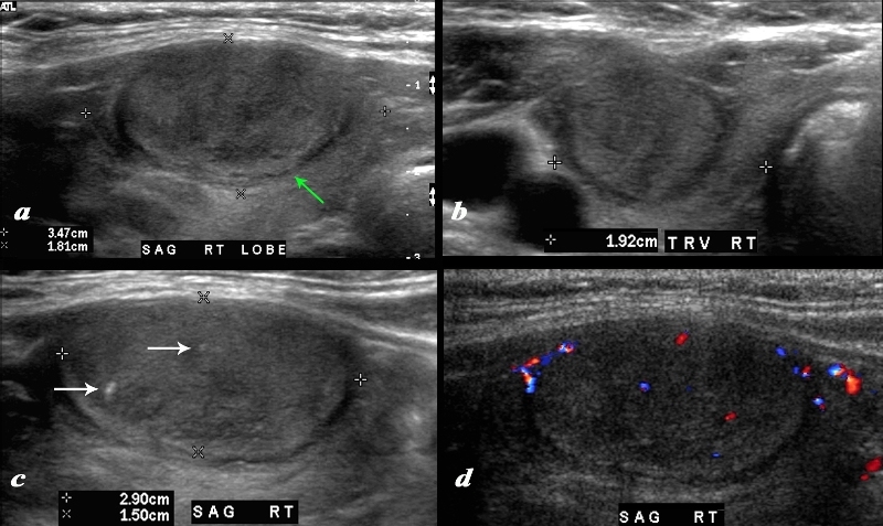
A large nodule in the thyroid occupies almost the entire right lobe (a). The nodule measures 2.9cms by 1.5cms. The gland is not enlarged and measures 3.5cms (craniocaudad), by 1.8cms (A-P) by 2.4cms (transverse). The nodule is almost isoechoic with normal thyroid but shows internal irregular areas of hypoechogenicity, regions of isoechogenicity, as well as microcalcifications (white arrows (c). There is irregularity of the border at the posterior aspect of the nodule green arrow a). The halo shows irregular borders in this region as well. Internal vascularity is minimal (d). The irregular surface is concerning for a malignant processes. The diagnosis in this patient was papillary carcinoma Courtesy Ashley Davidoff MD Copyright 2010 74909c02L.8 |
Combining Modalities
|
Normal Character by Thyroid Scan and Non Toxic Multiple Nodules by Ultrasound Strengths and Weaknesses of the Modalities Defining the Character of the Gland |
|
This is a thyroid scan from a 53 year old female with a clinical history of goiter. She was injected with Iodine -123 . 24 hours later radioiodine uptake was calculated at 31.8% (normal 8-35%). She was also injected with 14.2 mCi of 99mtechnetium pertechnitate.. The scan (a) shows an enlarged gland bilaterally with uniform uptake of the radioisotope in both lobes of the thyroid gland. The uptake in the thyroid gland is normal relative to the uptake in the submandibular and parotid glands. These findings are consistent with a diffuse goiter. The ultrasound on the other hand shows a normal sized gland with multiple nodules. The right lobe (c,d) measures 4.4cms (length) X 1.5cms (A-P) X 1.8cms (TRV) and the left lobe (f,g) measures 4.9cms (length) X 1.6cms (A-P) X 1.1cms (TRV) In the right lobe there is a nodule in the middle of the gland (b,c,d, e) that is relatively hypovascular that measures 1.8cms X 1.1 cms (d,e). Two 7mms nodules were also seen in the left lobe (f,g) Whereas the thyroid nuclear scan was able to accurately depict function, the ultrasound was more accurate in defining the exact size and nodular character of the gland. The diagnosis based on imaging therefore was a non toxic multinodular normal sized gland. Courtesy Alan Ashare MD and Ashley Davidoff MD Copyright 2010 97513c03.8L |

—