The Common Vein Copyright 2010
Introduction

CT Sensitivity to Small Calcifications and Positional Relationships |
|
The plain film (a), with magnified view (b) shows a large mass shifting and compressing the trachea (teal) to the right. The mass is seen on cross sectional CT as an irregularly enhancing mass connected to the thyroid gland on more cranial views. Its mass effect on the trachea narrowing it in the transverse dimension is well demonstrated. Two small but coarse calcifications are noted in the posterior aspect of the gland as 1mm white concretions. The findings are consistent with a retrosternal thyroid goiter Courtesy Ashley Davidoff MD Copyright 2010 31460c01.81s |
|
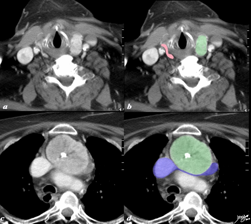
Retrosternal Goiter Compressing the Brachiocephalic Vein |
|
A large retrosternal goiter is seen extending down anterior to the aortic arch and brachiocephalic vein in this 86 year old female patient. In image a and b the atrophied left lobe of the thyroid is overlaid in pink, and the superior aspect of the goiter is overlaid in green. More inferiorly the goiter with central chunky calcification is noted compressing on the brachiocephalic vein (blue) There is mild diffuse almost homogeneous enhancement. The tracheobronchial tree is not affected by this goiter The findings are consistent with a calcified retrosternal thyroid goiter Courtesy Ashley Davidoff MD Copyright 2010 77978c.8s |
|
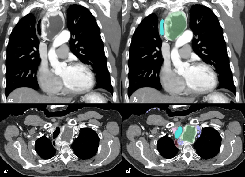
Retrosternal Goiter with Central Avascularity and Compressing the Brachiocephalic Vein |
|
A large retrosternal goiter is seen extending down anterior to the top aortic arch is noted in this 74 year old female patient. In image a and b the visualized portion of the goiter measures 8.8cms, has a central avascular component and an enhancing rind. The goiter is overlaid in green and seen displacing the trachea (teal blue) to the right. On the axial cuts the goiter with central avascularity is again noted compressing on the trachea and pushing it to the right (teal blue) the esophagus is also pushed rightward. The brachiocephalic vein (blue) is compressed and there are venous varicosities both on the left and posteriorly on the right of the goiter. Anterior chest veins are also prominent. The findings are consistent with a retrosternal thyroid goiter Courtesy Ashley Davidoff MD Copyright 2010 94516c.8s |