
The Common Vein copyright 2010
Introduction
In the Coronal Plane
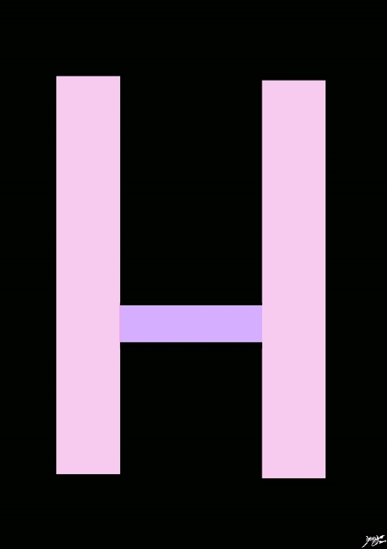
“H” shaped gland in the Coronal Projection |
|
The thyroid gland is an H shaped gland consisting of two vertical limbs called left and right lobes and a horizantal band connecting the two limbs called the isthmus Courtesy Ashley Davidoff MD copyright 2010 all rights reserved 94460b01.81s |
|
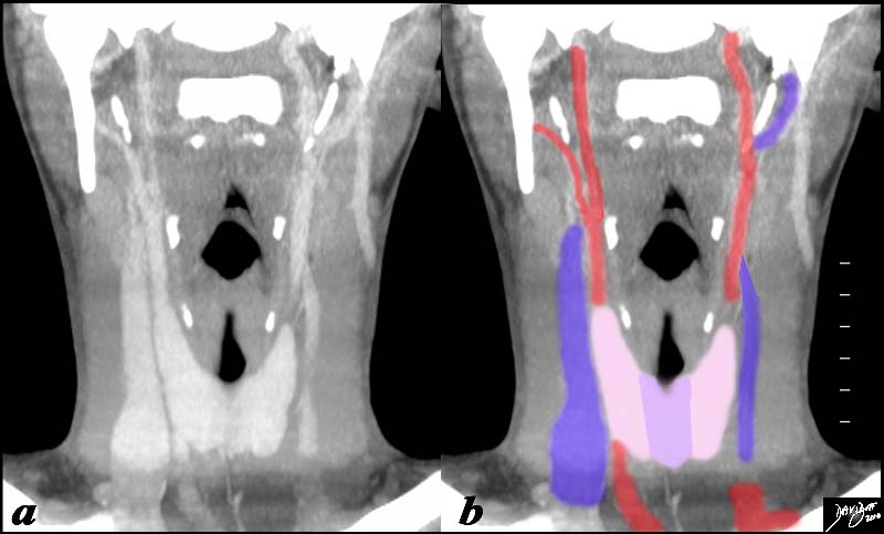
Coronal Reconstruction – CT scan – The Thyroid |
|
The CT scan of the neck shows the thyroid (pink) as a central structure surrounded by the great vessels including the internal jugular veins (blue) and the carotid arteries, (red) and the airway (black) Courtesy Ashley Davidoff MD copyright 2010 all rights reserved 93816c09b01.81s |
In the Transverse Plane
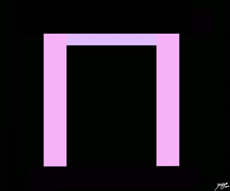
An invertyed “U” or an “n” |
|
The thyroid gland in the transverse view looks like an inverted “U” or an “n” with a right and left lobe represented in the as long and relatively wide limbs (pink) and the istmus represented asd a thinner and shorter lim connecting the two lobes (pale mauve) Courtesy Ashley Davidoff MD copyright 2010 all rights reserved 94883a.82s |

The Thyroid in Trasverse Section |
|
The axial CTscan of the neck k shows the thyroid gland with the right and left lobes (pink) and the isthmus in light purple. It is a central structure surrounded by the great vessels including the internal jugular veins (blue) and the carotid arteries, (red) and the airway (black) Courtesy Ashley DAvidoff MD copyright 2010 all rights reserved 30491c03.8s |
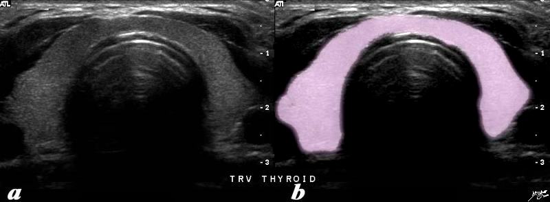
Transverse Scan using Ultrasound |
|
The transverse ultrasound of the thyroid glandshows the asymmetric symmetry of the lobes. In this cut the right lobe is slightly larger than the left and the isthmus consists of a narrow band that is only about 5mms thick. Courtesy Ashley DAvidoff MD copyright 2010 all rights reserved 77510c02.81s |