The Common Vein Copyright 2011
Introduction
The thyroid lies on the anterior side of the neck, against and around the trachea, and reaches posteriorly to touch the esophagus. It is anchored to the laryngoskeleton, and thus swallowing causes it to move up as the pharynx rises. It lies just below the cricoid cartilage. The cricoid cartilage is the landmark for the isthmus cartilage.
Relationships – Applied Anatomy
Anatomical relationships of the thyroid gland have clinical relevance; the recurrent laryngeal nerves lie in a groove between the lateral edges of the thyroid and the trachea behind the gland. In addition there are two pairs of parathyroid glands that are usually located in the back of the thyroid in the upper and middle portion of the lobes, but may be located within the gland.
The right recurrent laryngeal nerve originates from the vagus nerve, and after looping around the subclavian artery, it ascends behind the right lobe of the thyroid.
The left recurrent laryngeal nerve comes from the left vagus nerve, and after looping posteriorly around the aortic arch, it and ascends in the tracheoesophageal groove posterior to the left lobe of the thyroid.
The thyroid is also draped around the trachea and the esophagus posteriorly. All these structures can be affected by thyroid diseases like thyroid enlargement causing compression, or thyroid malignancies causing invasion. Surgical interventions may result in injury to these organs due to their close proximity.
|
Coronal Reconstruction – CT scan – The Thyroid |
|
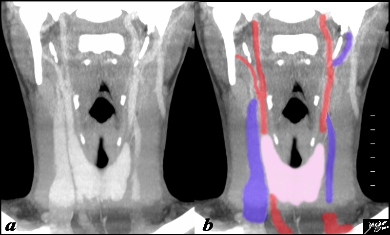
The CT scan of the neck shows the thyroid (pink) as a central structure surrounded by the great vessels including the internal jugular veins (blue) and the carotid arteries, (red) and the airway (black). Courtesy Ashley Davidoff MD copyright 2010 all rights reserved 93816c09b01.8s |
|
Position of the Thyroid in relation to the Carotid Arteries |
|
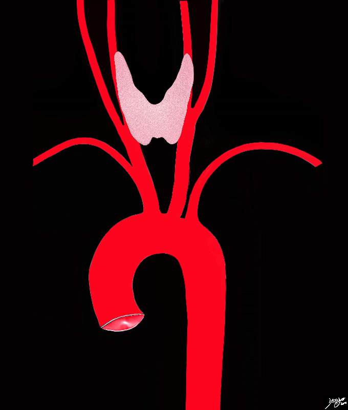
The thyroid gland lies anterior to the internal carotid arteries Courtesy Ashley Davidoff MD Copyright 2010 All rights reserved 47680d05.8kb03b.8s |
|
Transverse CT scan Showing Position and Relations |
|
The axial CT demonstrates the central position of the thyroid in the neck and its relations. Anteriorly a group of muscles called the strap muscles (tan) separate the thyroid gland from the skin. The thyroid surrounds the trachea (teal) on its anterior and lateral borders. The esophagus (darker pink) lies posterior to the trachea and medial to the posterior border of the lobes of the thyroid. The common carotid arteries lie posterior to each of the lobes, while the internal jugular veins lie lateral to the lobes. The prevertebral muscles (light brown) and vertebral arteries (bright red) form posterior relations of the thyroid. Courtesy Ashley Davidoff MD copyright 2010 all rights reserved 30491c02b02L.9s |
|
Retrosternal Goiter |
|
The plain film (a), with magnified view (b) shows a large mass shifting and compressing the trachea (teal) to the right. The mass is seen on cross sectional CT as an irregularly enhancing mass connected to the thyroid gland on more cranial views. Its mass effect on the trachea narrowing it in the transverse dimension is well demonstrated. The findings are consistent with a retrosternal thyroid goiter Courtesy Ashley Davidoff MD Copyright 2010 31460c01.8s |


