The Common Vein Copyright 2010
Definition
A follicular adenoma is a common benign tumor structurally characterized by a well large single, well circumscribed lesion most commonly non functioning.
Clinically they are painless and may be an incidental finding as a large (usually 3cms or more) painless
Diagnosis is initially based on an ultrasound which identifies a large well defined solid mass, and fine needle aspiration suggests the diagnosis but requires surgical removal for full pathological examination and for exclusion of carcinoma.
Adenomas are usually cold nodules since they usually take up less radioactive iodine than normal surrounding gland. About 10% of cold nodules are malignant.
Pathologically the largest clue to malignancy is capsular invasion.
Treatment is by surgical removal .

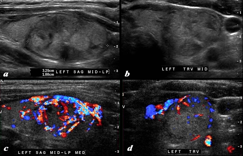
Domionant MAss – Indeterminate Biopsy Showed Follicular Adenoma |
|
This 37 year old female presents with single nodule in the left lobe of the thyroid. Ultrasound of the mass in sagittal (a) and transverse view (b) reveals a complex mass with isoechoic and hypoechoic nodular components. The normal thyroid tissue is seen superiorly in image a and medically in image b. In the sagittal view there also appears to be a small shadowing macrocalcification (arrow). The halo is seen partly around the superior aspect on the sagittal view and shows no definite irregularity. The Doppler study shows mostly a peripheral pattern but some prominent central vessels are shown. There are no definite criteria to suggest malignant disease but in view of the size, age and vascular pattern biopsy was performed and showed a follicular adenoma. Courtesy Ashley Davidoff MD Copyright 2010 95654c02.8 |

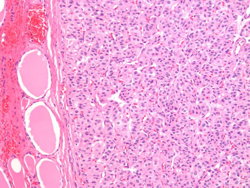
Microfollicular Adenoma H&E 20X |
|
The histological section at 20X magnification using H&E stain shows a follicular adenoma with microfollicles. The microfollicles appear as glandular structures with well defined lumens that are either empty. The size of the follicles are very similar. To the left of the image you can see the collagen capsule, and normal follicles outside the capsule. Image Courtesy Ashraf Khan MD. Department of Pathology, University of Massachusetts Medical School. 99396.8 |
